Sacroiliac Joint Injections, Lateral Branch Blocks
Revised February 4, 2015, by
Maxim S. Eckmann, MD
Director, Pain Medicine
Department of Anesthesiology
University of Texas Health Science Center at San Antonio
San Antonio, TX
Original Authors
Ronald Wasserman, MD
Chief, Division of Pain Medicine
Director, Back and Pain Center
Chad M. Brummett, MD
Director, Adult Pain Research
Division of Pain Medicine
Department of Anesthesiology
Division of Pain Medicine
University of Michigan Health System
Ann Arbor, MI
Acknowledgements: The authors would like to thank Dr. Kevin K. Tremper, Ph.D., M.D. (Professor and Chairman, Department of Anesthesiology, University of Michigan, Ann Arbor, MI) for guidance and support. We also thank Steven P. Cohen, M.D. (Associate Professor, Johns Hopkins University, Department of Anesthesiology and Critical Care, Baltimore, MD) for assistance.
Introduction
Sacroiliac joint (SIJ) syndrome is defined as pain originating from the SIJ due to degeneration or altered joint mobility. The degree to which low back pain is caused by pathologic conditions or dysfunction of the SIJ has been discussed for many decades. The SIJ is one of the many potential differential diagnoses in patients presenting with low back, buttocks and/or leg pain. A recent analysis of interventional pain procedure utilization in the Medicare population shows that sacroiliac joint injection is among the most common of interventional pain procedures, behind epidural and facet interventions, and experiencing rapid growth among a variety of practitioners.[1] This chapter will describe the epidemiology, anatomy, pathophysiology, diagnosis, and treatment of SIJ syndrome.
Epidemiology
The SIJ has been implicated as the primary source of pain in 10% to 26.6% of cases with suspected SIJ pain utilizing controlled comparative local anesthetic blocks on patients based on International Association for the Study of Pain (IASP) criteria.[2-6] The sacroiliac joint was widely considered a major source of back pain.[3][7-8] until Mixter and Barr in 1934 described disc herniation as a source of pain in the lumbar spine.[9] The evidence supporting the SIJ as a mechanical pain generator (in the absence of demonstrable inflammatory joint disease) was largely empirical, however, being mostly derived from successful treatment of patients with suspected sacroiliac joint pain.[5] Moderate degenerative joint changes occur as early as the 3rd decade of life.[10]
International Association for the Study of Pain (IASP) Diagnosis of Sacroiliac Joint Pain (Syndrome)
- Pain in the region of the sacroiliac joint
- Reproduction of concordant pain by physical examination techniques that stress the joint
- Elimination of pain with intra-articular injection of local anesthetic
(From Merskey H, Bogduk N: Classification of chronic pain, Descriptors of Chronic Pain Syndromes and Definitions of Pain Terms, 2nd Edition. Edited by Merskey H, Bogduk N. Seattle, IASP Press, 1994, pp. 206-207.)

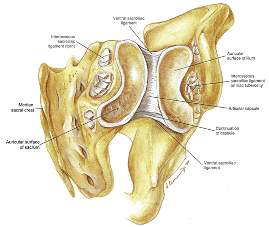
Figure 1. Posterior view of an opened SI joint illustrating the kidney-shaped lateral portion of the sacrum and medial aspect of the ilium, along with the articular capsule anteriorly and the interossesous sacroiliac ligaments (torn away) posteriorly.
(From Cramer GD, Chae-Song R: The Sacrum, Sacroiliac Joint, and Coccyx, Spine, Spinal Cord, and ANS, Second Edition. Edited by Cramer GD, Darby SA. St. Louis, Elsevier Mosby, 2005, pp 308-336)

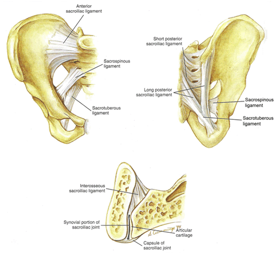
Figure 2. Figure showing the ligaments of the SI joint in the A. Anterior view, B. Posterior view and C. Horizontal section. Figure C illustrates that an articular capsule lines the anterior aspect of the joint and the posterior aspect is covered by the interosseous sacroiliac ligament.
(From Cramer GD, Chae-Song R: The Sacrum, Sacroiliac Joint, and Coccyx, Spine, Spinal Cord, and ANS, Second Edition. Edited by Cramer GD, Darby SA. St. Louis, Elsevier Mosby, 2005, pp 308-336.)
Anatomy
The SIJ is a large kidney-shaped, diarthrodial synovial joint of variable size, shape and contour. It is formed by the lateral portion of the sacrum and medial aspect of the ilium (Figure 1). The cephalad limb of this surface is oriented posteriorly and superiorly, and the caudad limb is oriented posteriorly and inferiorly. An articular capsule lines the SIJ’s anterior aspect, whereas the SIJ’s posterior aspect is covered by the interosseous sacroiliac ligament (Figure 2).[11-13] Thus, the majority of the true joint space is limited to the anterior portion; however, there is a small part of the synovial joint space that extends to the posterior and inferior portion of the SI apposition. This inferior portion of the joint is most commonly accessed for intra-articular injection. The joint’s stability comes not only from a complex network of surrounding ligaments, but also from a series of interlocking elevations and depressions, which become more irregular with age and can be altered with trauma. Stability can be reduced in conditions that promote ligamentous laxity.
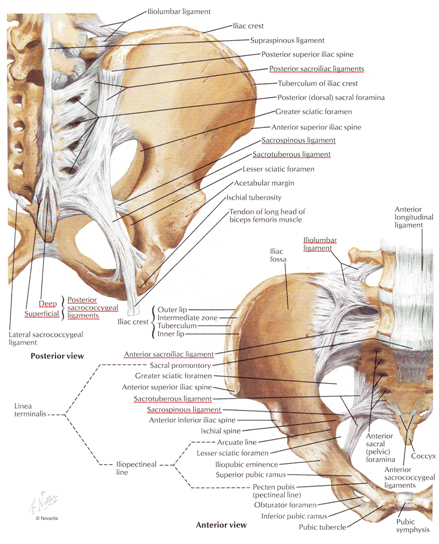
The SIJ is supported by an intricate system of ligaments. The interosseous ligament is the strongest ligament in the body and, along with other key ligaments, serves to tightly connect the sacrum and ilium together (see Figures 1 and 2C).[11] It acts to stabilize the joint and serves as the posterior joint border. The long (vertically oriented) and short (horizontally oriented) posterior sacroiliac ligaments cover the interosseous ligament (Figure 2B). The anterior surface (pelvic side) of the SIJ is covered by the anterior (ventral) sacroiliac ligament (Figure 2A), which provides less support than the posterior ligaments. The sacrotuberous, sacrospinous and iliolumbar ligaments also provide stability to the SIJ (Figure 3).[13] There are many other ligaments and muscles (a few of which include gluteus maximus and medius, piriformis, biceps femoris muscles) that contribute to the stability of the joint and restrict excessive motions during activities, such as walking, standing and sitting.

Figure 3. Figure illustrating the ligaments of the SIJ in both a posterior (top) and anterior (bottom) view. Note the long and short posterior ligaments, the sacrospinous, and sacrotuberous ligaments in the posterior view. Note the anterior sacroiliac ligament, iliolumbar ligament, the sacrospinous, and sacrotuberous ligaments in the anterior view.
(From Netter FH: Bones and Ligaments of Pelvis, Atlas of Human Anatomy, Second Edition. Edited by Netter FH. East Hanover, Hoechstetter Printing Company Inc, 1997, pp 331.)
The innervation of the SIJ has been a point of great interest in recent years due to the increase in interventional procedures designed to lesion the afferent sensory innervation of the posterior joint.[14-17] Both nociceptors and mechanoreceptors innervate the joint capsule and ligaments.[18-21] Many experts agree that the posterior portion of the joint comes from the lateral branches of L4-S3 dorsal rami, with some arguing that L3 and S4 may also contribute.[3][15-16][22]The lateral branch nerves then divide resulting in multiple smaller branches, some of which enter the SIJ and others do not. The location and number of the lateral branch nerves innervating the SIJ is highly variable (Figure 4).[23] The innervation of the anterior SIJ is unclear, as is the relative importance of the anterior joint innervation in SIJ pain. The lateral sacral arterial branches from the internal iliac artery and the median sacral artery provide the blood supply to the SIJ.

Figure 4. Figure illustrating the fluoroscopic anatomy of the lateral branch nerves at S1 (A), S2 (B), and S3 (C). Notice the variability in the course of the lateral branch nerves not only from level to level but also on the opposite side at the same levels.
(From Yin W, Willard F, Carreiro J, Dreyfuss P: Sensory stimulation-guided sacroiliac joint radiofrequency neurotomy: technique based on neuroanatomy of the dorsal sacral plexus. Spine 2003; 28: 2419-25)
Function
The SIJ provides stability to the region between the spine and lower extremities, while allowing for slight mobility to occur between the sacrum and ilium. It rotates about in all three axes, although the movements are very small and difficult to measure.[3] Various motions of the SIJ have been proposed, including gliding, tilting, rotation, and translation. Even though the precise nature of these motions is unclear, SIJ function may be affected by dynamic changes of the lumbar spine, hips and symphysis pubis.[24] Even though there appears to be no muscle specifically designed for movement of the SIJ, approximately 40 muscles can influence this joint of which the most important are the erector spinae, quadratus lumborum, multifidus, lumborum, iliopsoas, rectus abdominis, gluteus maximus, and piriformis muscles.
As we age, the stability of the SIJ increases, while the mobility decreases. Ligaments maintain joint stability until puberty after which point interlocking bone begins to form due to joint irregularities and crevice formation. Degenerative changes begin to form on the iliac side as early as the third decade in men and these are manifested by increased joint irregularity. Degenerative changes do not affect the sacral side until the fourth decade, and accelerated changes occur in men after the age of fifty. Fibrous ankylosis may develop at this age. The SIJ is locked in place near the eighth decade due to total fibrous degeneration resulting in no movement at this joint. There are no significant differences in the degree or types of joint motion in patients with SIJ pain and asymptomatic volunteers.[25]
Pathophysiology
The pathophysiology of SIJ pain is complicated by the many structures associated and the multitude of potential predisposing factors. Pain can come from intra-articular (arthritis, infections) and extra-articular (fractures, ligament injury, and myofascial) sources. A number of pathologic conditions that can cause pain in the SIJ are infection, inflammation, degeneration, trauma, metabolic changes, and tumor.[3][5] Compressive forces and stretching that go beyond that which the SIJ can withstand can cause injury to the bony structures and ligaments. This can be from bending, sitting, lifting, arching or twisting movements of the spine, or, from weight bearing forces associated with running or jumping. Even driving for long periods over rough terrain or poor suspension can cause SIJ pain because the bouncing reaches the SIJ directly without going through any other joint. Years of simple repetitive motions can wear the joint.
There are some conditions that predispose patients to SIJ pain. One of the most common is pregnancy.[26] During pregnancy there is increased mobility of the SIJ related to the hormone relaxin, which results in relaxation of the ligaments holding the SI joints together. This increased motion in the joints along with the additional weight and altered gait associated with pregnancy places additional stress on the SI joints, which can lead to pain. Other patients can be affected by leg length discrepancy and pain in the ankle, knee or hip resulting in an abnormal walking pattern. These all lead to increased stress on the SIJ. Lumbar spine surgery can also place extra joint stress due to increased force transmission, as well as for reasons unrelated such as SI ligament weakening and/or surgical violation of the joint cavity during iliac graft bone harvest and postsurgical hypermobility.[27]
The term SIJ dysfunction is used to explain pain from a SIJ that exhibits no demonstrable lesion, but which is presumed to have some type of biomechanical disorder that causes pain. Some believe that this condition can be diagnosed by detecting biomechanical abnormalities on physical examination.[28]
Diagnosis
History and Physical Examination
There are no absolute historical, physical, or radiological features to provide definitive diagnosis of SIJ pain.[3] The IASP criteria are noted in Table 1.[2] While the diagnostic criteria seem simple and clear, there are a multitude of other disorders (lumbar facet pain, intervertebral disc disease, spinal stenosis, myofascial pain, intra-articular hip pathology) that can present with similar symptoms and findings. In some patients, the etiology of the pain can be multifactorial combining any of the other noted pathologies with SIJ pain.
The variability and complexity of SIJ innervation noted in the anatomy section above make pain referral patterns highly variable. Pain referral patterns are one of the main historical findings that clinicians use to determine suspected cases of SIJ pain, but they cannot be used to make a specific diagnosis.[3] The most common site of pain is the ipsilateral buttocks (94%). Other sites in descending order include lower lumbar region (72%), lower extremity (50%), groin (14%), upper lumbar region (6%), and abdomen (2%).[29] Younger patients can describe pain below the knee, which can appear to be in an L5/S1 dermatomal distribution. The back/buttock pain is most commonly seen below the level of the L5 spinous process.[6][28-29] Maximal pain below L5 in combination with the posterior superior iliac spine or local tenderness just medial to the posterior superior iliac spine had the highest positive predictive value for the SIJ as a source of pain.[30]
Many physical examination tests have been studied in SIJ pain. Palpation over the caudad portion of the joint is commonly reported as painful in patients with SIJ pain. Unfortunately, most of the provocative maneuvers stress other potential pain structures, including the intervertebral discs, facet joints, muscles, and hip at the same time and therefore do not isolate the SIJ alone.[31] Common provocative physical examination tests include:
- Patrick's test (also known as the FABER test): see Figure 5
- Gaenslen's test: see Figure 6
- Yeoman's test (also known as the extension test): see Figure 7
- Gillet's test: see Figure 8
- Fortin's finger test: Ask the patient to point to the area of pain using only one finger. If the site is within 1 cm of the PSIS the test is considered positive.

Figure 5. Patrick's Test (also known as the FABER Test). The patient is supine and the affected hip is placed in Flexion, Abduction, and External Rotation. The ankle of the affected side is placed on the contralateral knee and then downward pressure is applied on the medial portion of the knee of the affected side, while counterpressure is provided on the contralateral anterior superior iliac spine. Pain in the buttock on the contralateral side of the flexed/abducted/externally rotated hip represents a positive test, while back and ipsilateral groin pain are less specific and can be signs of other pathology in the low back or hip structures, respectively.
(From Benzon HT: Pain Originating from the Buttock: Sacroiliac Joint Dysfunction and Piriformis Syndrome, Essentials of Pain Medicine and Regional Anesthesia, Second Edition. Edited by Benzon HT, Raja SN, Molloy RE, Liu SS, Fishman SM. Philadelphia, Elsevier Churchill Livingstone, 2005, pp 356-365.)

Figure 6. Gaenslen's Test. The patient is supine with the hip and knee of the unaffected limb flexed. The examiner then allows the affected limb to slowly fall off the side of the bed to passively extend the hip. The test is positive if pain is produced along the SIJ of the affected limb.
(From Benzon HT: Pain Originating from the Buttock: Sacroiliac Joint Dysfunction and Piriformis Syndrome, Essentials of Pain Medicine and Regional Anesthesia, Second Edition. Edited by Benzon HT, Raja SN, Molloy RE, Liu SS, Fishman SM. Philadelphia, Elsevier Churchill Livingstone, 2005, pp 356-365.)

Figure 7. Yoeman's (Extension) Test. The patient is placed prone. The examiner puts one hand over the anterior aspect of the knee of the affected limb and elevates it to passively extend the hip while simultaneously applying downward pressure with the other hand over the contralateral iliac crest. The test is positive if pain is provoked along the SIJ of the affected limb.
(From Loomba D, Mahajan G: Sacroiliac Joint Pain, Current Therapy in Pain. Edited by Smith HS. Philadelphia, Saunders Elsevier, 2009, pp 354-363.)

Figure 8. Gillet's Test. With the patient standing the examiner places one thumb on the second sacral spinous process, while the other thumb is placed on the posterior iliac spine. Have the patient maximally flex the hip. The posterior superior iliac spine (PSIS) on the ipsilateral side moves inferior to the S2 spinous process in a normal SIJ. With a dysfunctional or fixed SIJ the PSIS remains at the level of the S2 spinous process or moves superior to the sacrum.
(From Benzon HT: Pain Originating from the Buttock: Sacroiliac Joint Dysfunction and Piriformis Syndrome, Essentials of Pain Medicine and Regional Anesthesia, Second Edition. Edited by Benzon HT, Raja SN, Molloy RE, Liu SS, Fishman SM. Philadelphia, Elsevier Churchill Livingstone, 2005, pp 356-365)
Radiological Imaging
There are no radiological studies that provide a definitive diagnosis of SIJ syndrome. The incidence of degenerative SIJ changes on X-ray is approximately 25% in asymptomatic individuals > 50 years old.[32] By assuming that those patients who responded positively to SIJ injection had pain originating from the SIJ a number of studies have been done to assess the sensitivity and specificity of a variety of imaging modalities. Bone scan has poor sensitivity ranging from 13%[33] to 46%[34] but appears to have good specificity (90%).[34] Computed tomography (CT) has a sensitivity of 57.5% and a specificity of 69%.[35] Single-photon emission computed tomography (SPECT) has a poor sensitivity of 9.1%.[29] While MRI findings such as bone marrow edema can be reasonably useful for diagnosing sacroilitis in spondyloarthropathies[47], studies assessing the efficacy of MRI for SIJ syndrome are inconclusive.

Figure 9. Intra-articular Sacroiliac Joint Injection.

Figure 10. After placement of the needle into the SIJ, contrast dye is injected. Contrast can be seen outlining the inferior portion of the joint in this fluoroscopic image. Some extra-articular spread is also noted, which is very common.
Diagnostic Blocks
There is no single test or intervention that is sensitive or specific for the diagnosis of pain originating from SIJ syndrome. Due to the lack of sensitivity and specificity of history, physical examination, and other testing modalities, intra-articular sacroiliac joint blocks have become the mainstay of diagnosis and treatment. Although SIJ blocks may be better than other measures, there is still potential for false-positive (estimated at about 20%[36]) and false-negative responses. Some have recommended the use of lumbar medial branch and sacral lateral branch diagnostic blockade with local anesthetic as an alternative means to diagnosis;[14][22] however, the failure to block the innervation of the anterior joint, the variable location of the lateral branches of the sacral roots, and the spread of local anesthetic to other pain-generating structures make this approach even less attractive than a diagnostic intra-articular SIJ block. There is no accepted gold-standard for diagnostic blockade of the SIJ, thereby making an accurate estimate of the prevalence of SIJ pain and false-negative and false-positive blocks impossible.
Injection specifically into the SIJ can be challenging, and extra-capsular spread is extremely common.[30] Technical challenges may be greatest in elderly patients with significant degeneration and bony changes.[3] Intra-articular SIJ injections are performed using fluoroscopic (Figures 9 and 10) or more rarely CT, guidance. Blind, landmark guided injections have poor success rates, with intra-articular injection accomplished in less than 25% of the cases.[37] Unlike diagnostic medial branch blocks (MBBs) for lumbar facet pain, the intra-articular injection of the SIJ usually incorporates steroid along with the local anesthetic as a treatment in common clinical practice. Therefore, comparative local anesthetic blocks or placebo-controlled blocks are less commonly performed for SIJ pain than for lumbar facet joint pain. However, if radiofrequency denervation is planned as a long-term treatment for SIJ pain, a similar diagnostic paradigm can be applied for controlled lateral blocks.
Treatment
Conservative Management
While the use of interventional techniques for SIJ pain have increased in recent years, the use of conservative therapies prior to or in conjunction with intervention is warranted. Joint mobilization can be considered for cases of restricted joint mobility and associated pain provided there are no contraindications like osteoporosis. Joint stabilization belts can help in cases of pain associated with joint laxity, for example in pregnancy.
Pharmacotherapy
The pharmacotherapeutic treatment for SIJ pain is comparable to other pain conditions, such as facet pain.[38] There are no randomized, controlled trials for the therapy of SIJ pain. Non-steroidal anti-inflammatory medications are the best early option. Some patients will derive benefit from acetaminophen without the potential for significant side effects. Adjunctive medications, including membrane stabilizers (i.e. gabapentin), tricyclic antidepressants, and serotonin-norepinephrine reuptake inhibitors are reasonable options. The authors do not recommend the regular use of opioids for SIJ pain.[3]
Physical and Occupational Therapy
A physical therapy evaluation should include evaluation for any obvious anatomic abnormalities, such as leg length discrepancy. Relative position and mobility between the two SIJs can be assessed, correlated with pain, and possibly corrected with manual therapy and stretching.Postural and gait changes can be beneficial. Hyperactivity of the gluteus muscle can occur in some patients, and therapy targeting gluteal muscle can improve pain in some patients. [39]
Psychotherapy
As with all pain conditions, untreated psychiatric comorbidities will adversely affect the patient’s response to virtually all treatments and interventions. Even patients without premorbid psychopathology can have significant difficulty due to pain and loss of function from sacroilitis and SIJ syndrome. Cognitive-behavioral approaches can reduce fear-avoidance behaviors.
Intra-articular Joint Injections
As was noted in the diagnostic section above, SIJ joint injections serve as both a diagnostic and therapeutic measure. There have been a number of studies demonstrating benefit from the intra-articular injection of local anesthetic and steroid. Fully assessing the outcomes of these studies, as with other interventions to treat SIJ pain, is difficult due to the lack of a measure to accurately diagnose SIJ pain. The majority of the studies have been prospective, observational trials, which generally show good benefit.[40] There are limited data to support a long-term benefit from intra-articular steroids; however, short-intermediate-term (months) benefit has been shown.[3][36] In a randomized, placebo-controlled, double blind study, Maugars et al.[41] studied patients with SIJ pain diagnosed through clinical and radiological measures and demonstrated a significant improvement in relief with the addition of steroid when compared with local anesthetic only. Non-responders strongly suspected to have pain originating from the SIJ may benefit from a repeated injection.[42] Periarticular injections with steroid have also been found to provide pain benefit in patients with suspected SIJ pain.[43-44]
There are multiple descriptions for the technique for SIJ injection. Figures 9 and 10 demonstrate a 22-gauge needle in the caudad portion of the SIJ with contrast outlining the joint. After confirming intra-articular joint placement with contrast dye spread and the firm feel of the needle placement within the joint, the local anesthetic and steroid mixture is injected. Usually no more than 1.5–2 ml is injected due to a small joint space. For a more detailed explanation of the technique of SIJ injection, we recommend the Atlas of Image-Guided Regional Anesthesia and Pain Medicine by James P. Rathmell[45] and the International Spine Intervention Society: Practice Guidelines-Spinal Diagnostic and Treatment Procedures.[46]

Figure 11. The target points for denervation of the lateral branches of L4 and L5 and the S1-S3 roots are noted in this figure. Some experts recommend an additional lesion at the lateral border of S4.
(From Cohen SP, Hurley RW, Buckenmaier CC, 3rd, Kurihara C, Morlando B, Dragovich A: Randomized placebo-controlled study evaluating lateral branch radiofrequency denervation for sacroiliac joint pain. Anesthesiology 2008; 109: 279-88)

Figure 12. (A) The wider lesion size of a water-cooled radiofrequency (RF) is noted. (B) Shows monopolar lesion size along the length of the active tip of the traditional RF needle. The clinical importance of the larger lesion size with water-cooled RF is suspected but unproven.
(From Cohen SP, Hurley RW, Buckenmaier CC, 3rd, Kurihara C, Morlando B, Dragovich A: Randomized placebo-controlled study evaluating lateral branch radiofrequency denervation for sacroiliac joint pain. Anesthesiology 2008; 109: 279-88)
Radiofrequency Denervation
Whereas most patients who receive intra-articular SIJ blocks will derive partial to substantial relief, there are some patients that fail to have prolonged benefit. There have been studies in recent years investigating the effects of lateral branch radiofrequency neurotomy of the lower lumbar and sacral region as a means to denervate the posterior SIJ. While some researchers have investigated the effects of diagnostic lateral branch blocks as a predictive measure of denervation, the complicated and unpredictable anatomy of the sensory innervation of the posterior SIJ make intra-articular SIJ injection with local anesthetic alone, or with steroid, more attractive.[14][16][22] In a well-designed, randomized, controlled trial, Cohen et al.[15] demonstrated that thermal radiofrequency lesioning of the L4 and L5 dorsal primary rami and water-cooled radiofrequency lesioning of the S1-S3 lateral branches provided significantly more pain relief at a 1-month follow-up when compared with the placebo group.
Many of the technical aspects of SIJ denervation are similar to denervation of the lumbar medial branches.[38] In fact, the L4 medial and lateral branches and primary dorsal ramus of L5 are completed in the same fashion. The lateral branches of S1-S3 are a more variable in their position, requiring broader lesioning. Most experts recommend three target points around the lateral portion of the sacral foramen as shown in Figure 11.[15] Water-cooled radiofrequency devices create a larger lesion (Figure 12); however, there are limited data to support additional efficacy when compared with traditional radiofrequency devices.
Surgery
There are no well-designed studies supporting the use of surgical stabilization for the treatment of degenerative SIJ disease and pain. SIJ fusion is sometimes done for instability and fractures, but again, there are limited outcome data.
Summary
The SIJ is a common source of pain in the low back, buttocks, hip, and leg. Accurate diagnosis is difficult due to the lack of a sensitive and specific test; however, intra-articular SIJ injections are the most common current means of diagnosis. Treatment requires a multidisciplinary approach.
References
- Manchikanti L, Falco FJ, Singh V, Pampati V, Parr AT, Benyamin RM, Fellows B, Hirsch JA. Utilization of interventional techniques in managing chronic pain in the Medicare population: analysis of growth patterns from 2000 to 2011. Pain Physician. 2012 Nov-Dec;15(6):E969-82.
- Merskey H, Bogduk N: Classification of chronic pain, Descriptors of Chronic Pain Syndromes and Definitions of Pain Terms, 2nd Edition. Edited by Merskey H, Bogduk N. Seattle, IASP Press, 1994, pp 206-207.
- Cohen SP: Sacroiliac joint pain: a comprehensive review of anatomy, diagnosis, and treatment. Anesth Analg 2005; 101: 1440-53
- Cohen SP: Epidemics, evolution, and sacroiliac joint pain. Reg Anesth Pain Med 2007; 32: 3-6
- Hansen HC, McKenzie-Brown AM, Cohen SP, Swicegood JR, Colson JD, Manchikanti L: Sacroiliac joint interventions: a systematic review. Pain Physician 2007; 10: 165-84
- Schwarzer AC, Aprill CN, Bogduk N: The sacroiliac joint in chronic low back pain. Spine (Phila Pa 1976) 1995; 20: 31-7
- Hansen HC, Helm S, 2nd: Sacroiliac joint pain and dysfunction. Pain Physician 2003; 6: 179-89
- Slipman CW, Whyte WS, 2nd, Chow DW, Chou L, Lenrow D, Ellen M: Sacroiliac joint syndrome. Pain Physician 2001; 4: 143-52
- Mixter WJ, Barr JS: Rupture of the intervertebral disc with involvement of the spinal cord. N Engl J Med 1934; 211: 210-214
- Shibata Y, Shirai Y, Miyamoto M: The aging process in the sacroiliac joint: helical computed tomography analysis. J Orthop Sci 2002; 7: 12-8
- Cramer GD, Chae-Song R: The Sacrum, Sacroiliac Joint, and Coccyx, Spine, Spinal Cord, and ANS, Second Edition. Edited by Cramer GD, Darby SA. St. Louis, Elsevier Mosby, 2005, pp 308-336
- Loomba D, Mahajan G: Sacroiliac Joint Pain, Current Therapy in Pain. Edited by Smith HS. Philadelphia, Saunders Elsevier, 2009, pp 354-363
- Netter FH: Bones and Ligaments of Pelvis, Atlas of Human Anatomy, Second Edition. Edited by Netter FH. East Hanover, Hoechstetter Printing Company Inc, 1997, pp 331
- Cohen SP, Abdi S: Lateral branch blocks as a treatment for sacroiliac joint pain: A pilot study. Reg Anesth Pain Med 2003; 28: 113-9
- Cohen SP, Hurley RW, Buckenmaier CC, 3rd, Kurihara C, Morlando B, Dragovich A: Randomized placebo-controlled study evaluating lateral branch radiofrequency denervation for sacroiliac joint pain. Anesthesiology 2008; 109: 279-88
- Cohen SP, Strassels SA, Kurihara C, Crooks MT, Erdek MA, Forsythe A, Marcuson M: Outcome predictors for sacroiliac joint (lateral branch) radiofrequency denervation. Reg Anesth Pain Med 2009; 34: 206-14
- Ferrante FM, King LF, Roche EA, Kim PS, Aranda M, Delaney LR, Mardini IA, Mannes AJ: Radiofrequency sacroiliac joint denervation for sacroiliac syndrome. Reg Anesth Pain Med 2001; 26: 137-42
- Sakamoto N, Yamashita T, Takebayashi T, Sekine M, Ishii S: An electrophysiologic study of mechanoreceptors in the sacroiliac joint and adjacent tissues. Spine (Phila Pa 1976) 2001; 26: E468-71
- Vilensky JA, O'Connor BL, Fortin JD, Merkel GJ, Jimenez AM, Scofield BA, Kleiner JB: Histologic analysis of neural elements in the human sacroiliac joint. Spine (Phila Pa 1976) 2002; 27: 1202-7
- Yamashita T, Cavanaugh JM, el-Bohy AA, Getchell TV, King AI: Mechanosensitive afferent units in the lumbar facet joint. J Bone Joint Surg Am 1990; 72: 865-70
- Yamashita T, Minaki Y, Oota I, Yokogushi K, Ishii S: Mechanosensitive afferent units in the lumbar intervertebral disc and adjacent muscle. Spine (Phila Pa 1976) 1993; 18: 2252-6
- Dreyfuss P, Henning T, Malladi N, Goldstein B, Bogduk N: The ability of multi-site, multi-depth sacral lateral branch blocks to anesthetize the sacroiliac joint complex. Pain Med 2009; 10: 679-88
- Yin W, Willard F, Carreiro J, Dreyfuss P: Sensory stimulation-guided sacroiliac joint radiofrequency neurotomy: technique based on neuroanatomy of the dorsal sacral plexus. Spine (Phila Pa 1976) 2003; 28: 2419-25
- Calvillo O, Skaribas I, Turnipseed J: Anatomy and pathophysiology of the sacroiliac joint. Curr Rev Pain 2000; 4: 356-61
- Sturesson B, Selvik G, Uden A: Movements of the sacroiliac joints. A roentgen stereophotogrammetric analysis. Spine (Phila Pa 1976) 1989; 14: 162-5
- Berg G, Hammar M, Moller-Nielsen J, Linden U, Thorblad J: Low back pain during pregnancy. Obstet Gynecol 1988; 71: 71-5
- Ebraheim NA, Elgafy H, Semaan HB: Computed tomographic findings in patients with persistent sacroiliac pain after posterior iliac graft harvesting. Spine (Phila Pa 1976) 2000; 25: 2047-51
- Dreyfuss P, Michaelsen M, Pauza K, McLarty J, Bogduk N: The value of medical history and physical examination in diagnosing sacroiliac joint pain. Spine (Phila Pa 1976) 1996; 21: 2594-602
- Slipman CW, Jackson HB, Lipetz JS, Chan KT, Lenrow D, Vresilovic EJ: Sacroiliac joint pain referral zones. Arch Phys Med Rehabil 2000; 81: 334-8
- Fortin JD, Dwyer AP, West S, Pier J: Sacroiliac joint: pain referral maps upon applying a new injection/arthrography technique. Part I: Asymptomatic volunteers. Spine (Phila Pa 1976) 1994; 19: 1475-82
- Laslett M, Williams M: The reliability of selected pain provocation tests for sacroiliac joint pathology. Spine (Phila Pa 1976) 1994; 19: 1243-9
- Vogler JB, 3rd, Brown WH, Helms CA, Genant HK: The normal sacroiliac joint: a CT study of asymptomatic patients. Radiology 1984; 151: 433-7
- Slipman CW, Sterenfeld EB, Chou LH, Herzog R, Vresilovic E: The value of radionuclide imaging in the diagnosis of sacroiliac joint syndrome. Spine (Phila Pa 1976) 1996; 21: 2251-4
- Maigne JY, Boulahdour H, Chatellier G: Value of quantitative radionuclide bone scanning in the diagnosis of sacroiliac joint syndrome in 32 patients with low back pain. Eur Spine J 1998; 7: 328-31
- Elgafy H, Semaan HB, Ebraheim NA, Coombs RJ: Computed tomography findings in patients with sacroiliac pain. Clin Orthop Relat Res 2001: 112-8
- McKenzie-Brown AM, Shah RV, Sehgal N, Everett CR: A systematic review of sacroiliac joint interventions. Pain Physician 2005; 8: 115-25
- Rosenberg JM, Quint TJ, de Rosayro AM: Computerized tomographic localization of clinically-guided sacroiliac joint injections. Clin J Pain 2000; 16: 18-21
- Brummett CM, Cohen SP: Facet Blocks: Facet Joint Injections, Medial Branch Blocks, Rhizotomy., Raj's Practial Management of Pain, 4th Edition. Edited by Benzon HT, Rathmell JP, Wu CL, Turk DC, Argoff CE. New York, Elsevier-Mosby, 2006
- Mooney V, Pozos R, Vleeming A, Gulick J, Swenski D: Exercise treatment for sacroiliac pain. Orthopedics 2001; 24: 29-32
- Liliang PC, Lu K, Weng HC, Liang CL, Tsai YD, Chen HJ: The therapeutic efficacy of sacroiliac joint blocks with triamcinolone acetonide in the treatment of sacroiliac joint dysfunction without spondyloarthropathy. Spine (Phila Pa 1976) 2009; 34: 896-900
- Maugars Y, Mathis C, Berthelot JM, Charlier C, Prost A: Assessment of the efficacy of sacroiliac corticosteroid injections in spondylarthropathies: a double-blind study. Br J Rheumatol 1996; 35: 767-70
- Gunaydin I, Pereira PL, Fritz J, Konig C, Kotter I: Magnetic resonance imaging guided corticosteroid injection of sacroiliac joints in patients with spondylarthropathy. Are multiple injections more beneficial? Rheumatol Int 2006; 26: 396-400
- Luukkainen R, Nissila M, Asikainen E, Sanila M, Lehtinen K, Alanaatu A, Kautiainen H: Periarticular corticosteroid treatment of the sacroiliac joint in patients with seronegative spondylarthropathy. Clin Exp Rheumatol 1999; 17: 88-90
- Luukkainen RK, Wennerstrand PV, Kautiainen HH, Sanila MT, Asikainen EL: Efficacy of periarticular corticosteroid treatment of the sacroiliac joint in non-spondylarthropathic patients with chronic low back pain in the region of the sacroiliac joint. Clin Exp Rheumatol 2002; 20: 52-4
- Rathmell JP: Atlas of Image-Guided Intervention in Regional Anesthesia and Pain Medicine. Philadelphia, Lippincott Williams and Wilkins, 2005
- ISIS Practice Guidelines-Spinal Diagnostic and Treatment Procedures, 2nd ed. Bogduk N, International Spine Intervention Society, 2014.
- Aydin SZ, Maksymowych WP, Bennett AN, McGonagle D, Emery P, Marzo-Ortega H. Validation of the ASAS criteria and definition of a positive MRI of the sacroiliac joint in an inception cohort of axial spondyloarthritis followed up for 8 years. Ann Rheum Dis. 2012 Jan;71(1):56-60.