POCUS Spotlight: Rapid Ultrasound for Shock and Hypotension (RUSH) Protocol for the Perioperative Setting
Cite as: Douglas R, Pérez d’Empaire P, Santiago AS, et al. POCUS spotlight: rapid ultrasound for shock and hypotension (RUSH) protocol for the perioperative setting. ASRA Pain Medicine News 2026;51. https://doi.org/10.52211/asra110126.009.
POCUS Spotlight
Introduction
Postoperative hypotension is correlated with a 2.5 times greater risk of myocardial injury and patient mortality, an increased risk of delirium and longer hospital length of stay.1,2 Identifying etiology quickly matters.2 The rapid ultrasound for shock and hypotension (RUSH) protocol has a high degree of accuracy in differentiating etiologies of shock in emergency and trauma settings.3 In the attached videos and accompanying article, we present an amended version of the RUSH protocol for the everyday anesthesiologist, with a focus on troubleshooting perioperative hypotension.
Indications
Assessing for etiology of perioperative hypotension and shock.
Acquisition
Protocol
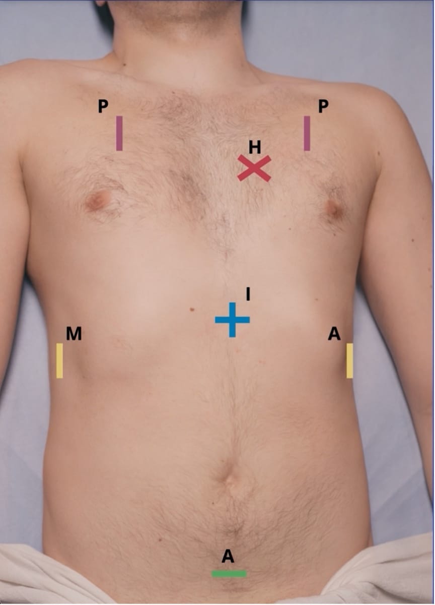
The RUSH protocol uses the HI-MAP acronym to outline the exam sequence: heart, inferior vena cava (IVC), Morrison’s pouch, abdominal views/focused assessment with sonography in trauma (FAST), and pulmonary scanning.4 There are 10 views in total. We start with four cardiac views: parasternal long axis (PLAX), parasternal short axis (SAX), apical four-chamber (A4C), and subcostal four-chamber (SC4C). Then, the protocol includes the IVC, Morrison’s pouch in the left upper quadrant (LUQ), the splenorenal view in the right upper quadrant (RUQ), and bladder views in the suprapubic area. Finally, the protocol incorporates anterior pulmonary views. We adapted the original RUSH protocol by excluding the scan to assess for an abdominal aneurysm, as this is an uncommon finding in the perioperative setting, and instead added the SAX and SC4C views. These views are especially useful for anesthesiologists because the SAX view quickly assesses left ventricle (LV) contractility and patient volume status, and the SC4C is easy to acquire in supine, intubated patients.
Patient Positioning/Preparation
The patient should be supine and can be rotated into a left tilt for the cardiac views.
Transducer Selection
To obtain the images, ideally use a phased-array probe for all views to improve efficiency, but you may change your probe selection for better resolution. The phased array is required for all the cardiac views. For abdominal views, you may use phased array and curvilinear probes, and for thoracic lung, you may use any probe. We suggest using the phased array probe when efficiency is important.
Interpretation and Medical Decision-Making:
We will use the HI-MAP algorithm (see Figure 1) to evaluate shock types: hypovolemic, distributive, obstructive, and cardiogenic (see Video 2).
POCUS Findings by Shock Etiology
| SHOCK TYPE | POCUS FINDINGS BY ETIOLOGY |
| Hypovolemic | Small LV cavity with preserved function, collapsible/ flat IVC, potential + FAST exam |
| Distributive | Hyperdynamic LV function, possible dynamic left ventricular outflow tract (LVOT) obstruction, normal/ low potential lung pathology |
| Obstructive (all subtypes have enlarged IVC, +/- B lines) |
|
| Cardiogenic | LV dysfunction, regional wall motion abnormalities, valvular disease, normal or dilated IVC, possible lung B lines |
Hypovolemic Shock
Hypovolemic shock is the most common type of shock encountered in the operating room.5 Hypovolemic shock results from a critical reduction in intravascular volume, initially compensated by increased vascular resistance but progressing to reduced cardiac output and hypotension. Common causes of hypovolemic shock encountered in the operating room include hemorrhagic and non-hemorrhagic causes. Overall, the heart and IVC POCUS exam has been shown to be reliable for ruling out or evaluating hypovolemic shock, with sensitivity and specificity of 95%.3
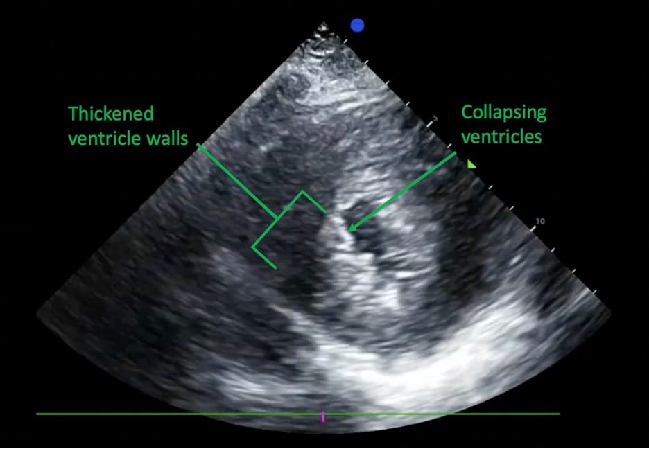
Heart: In hypovolemic shock, the left ventricular cavity often appears small, or you may see an underfilled left ventricle, with the cavity nearly collapsing at end-systole, commonly described as a “kissing ventricle.”6 This is best observed in the SAX view (see Figure 2). The heart may also be hyperdynamic, as further explained in our distributive shock section.
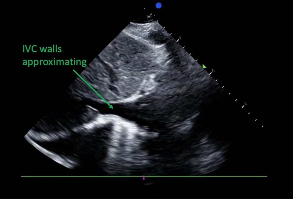
IVC: The IVC is often small and may show pronounced collapsibility in spontaneously breathing patients (see Figure 3), indicating volume responsiveness.7
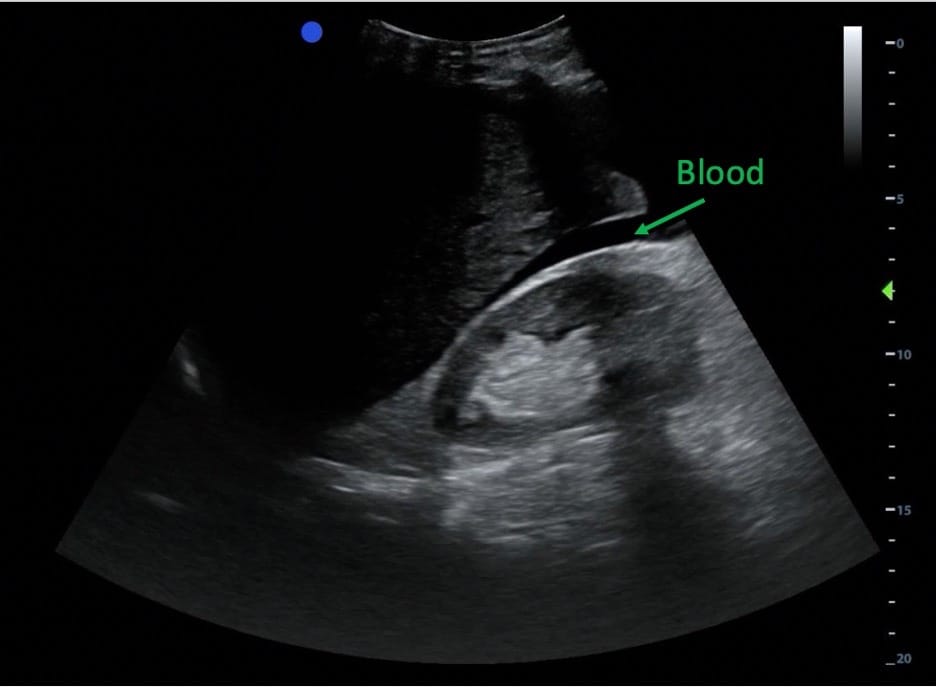
Morrison’s Pouch and Abdomen: If there is an intra-abdominal bleed in the peritoneum, you will likely observe free fluid in dependent areas, including Morrison’s pouch (see Figure 4), above the spleen (peri-splenic space), and behind the bladder.
Pulmonary: Pulmonary findings are usually minimal.
Distributive Shock
In distributive shock, the principal pathophysiological mechanisms are peripheral vasodilation, a lack of volume response, and tissue hypoxia, resulting in relative hypovolemia and, consequently, hypotension.8 Multiple etiologies can cause distributive shock: sepsis, anaphylaxis, neurogenic shock, adrenal insufficiency, high block level after spinal anesthesia, pancreatitis, and toxicities. It is the most common overall cause of shock among patients presenting to the hospital, accounting for more than half of shock cases.9
Heart: Although left ventricular contractility can be normal in some cases, the most common finding in distributive shock is a hyperdynamic heart.10 A hyperdynamic heart refers to how the heart contracts, not necessarily its contraction rate. It is characterized by vigorous (hypercontractile) wall motion and a low end-systolic volume, with an ejection fraction greater than 70%. This finding can be challenging to differentiate from hypovolemia. However, in hypovolemia, both end-systolic and end-diastolic volumes are smaller, whereas in distributive shock, only the end-systolic volume is smaller.11 In advanced POCUS, you can consider using the left ventricular outflow tract velocity-time integral (LVOT VTI) to help differentiate, with LVOT VTI potentially lower in hypovolemic shock.12 This was discussed in a recent POCUS Spotlight article in this newsletter.13 In the surgical setting, you may have a mixed hypovolemic and distributive shock etiology. In this situation, ultrasound can help you assess whether the heart appears empty vs. hyperdynamic and guide vasopressor therapy versus intravascular volume resuscitation. In sepsis, you may also see sepsis-induced cardiomyopathy, which presents with a moderately dilated LV and reduced ejection fraction (EF) and is often reversible with recovery within 7-10 days after the onset of sepsis.14
IVC: In distributive shock, the IVC usually looks small and collapsible. However, it may also be dilated if there is an obstructive pathology, ie, pericardial effusion.11
Morrison’s Pouch and Abdomen: In distributive shock, there shouldn’t be any appreciable abdominal pathology on ultrasound. However, when interrogating the lower thoracic cavities, you may see pulmonary findings.
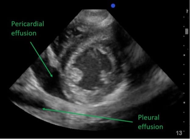
Pulmonary: If the lungs have an infectious septic etiology, you may see pathology such as pleural effusion (see Figure 5), exudate, empyema, or B lines, among other findings. In early sepsis, anaphylaxis, or a high spinal, you would expect the lungs to appear normal.15
Obstructive Shock
In the perioperative setting, obstructive shock is mostly caused by tension pneumothorax, cardiac tamponade, left ventricular outflow tract obstruction, or acute pulmonary embolism. All these conditions impair cardiac filling or outflow, thereby reducing effective cardiac output. In brief, during the HI-MAP sequence, you will see unique cardiac findings and a distended IVC, while Morrison’s pouch and other abdominal findings are normal. The pulmonary findings may vary based on the etiology.
Pneumothorax
A tension pneumothorax is a life-threatening emergency in which air enters the pleural space, causing mediastinal shift and obstructing venous return.
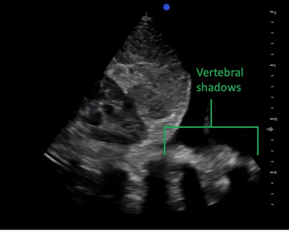
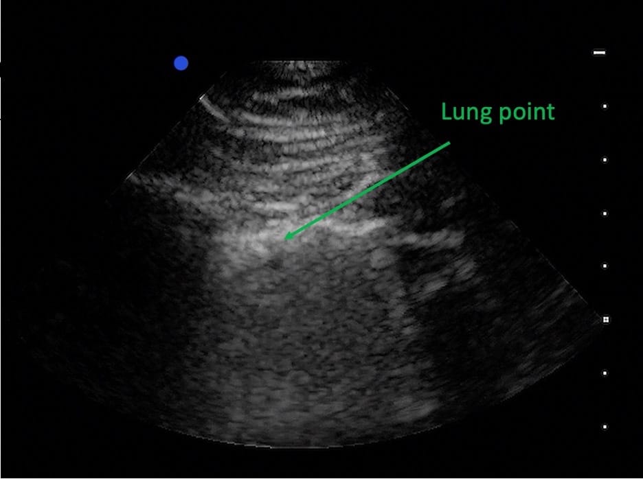
In pneumothorax, lung ultrasound may reveal findings consistent with a pneumothorax, such as absent lung sliding, absent lung pulse, and the presence of a lung point (see Figure 6).16 However, even though the lung point is pathognomonic and 100% specific for a pneumothorax,17 it won’t be found in a large tension pneumothorax. As noted in the Lichenstein paper where the lung point was first described, a lung point may be absent in a large pneumothorax where the lung is fully collapsed.18 Also of note, lack of lung sliding is not specific to pneumothorax and may also occur with lung bullae, pleurodesis, mainstem intubation, or lung collapse. A recent POCUS Spotlight article in the newsletter described lung findings in pneumothorax.19
Theoretically, in a tension pneumothorax, air compresses the heart and obstructs your view of it. But if you could see the heart, you would see evidence of an underfilled, hyperdynamic heart.
Cardiac Tamponade
Cardiac tamponade is primarily a clinical diagnosis that often presents with exertional dyspnea, hemodynamic instability, and distended neck veins with minimal respiratory variation, but classic physical findings occur in fewer than 40% of confirmed cases.20 Ultrasound is therefore valuable for confirming the diagnosis, with pericardial fluid appearing as an anechoic collection within the pericardial sac (see Figure 7); up to 50 mL of pericardial fluid can be physiologic, so assessment should include at least two cardiac views. In the PLAX view, fluid posterior to the aorta suggests a pleural effusion, whereas fluid anterior to the aorta indicates a pericardial effusion.
Additional sonographic features suggestive of cardiac tamponade include right atrial and right ventricular systolic collapse and a heart that may appear to swing within a large pericardial effusion. Right atrial collapse is the earliest ultrasound sign of cardiac tamponade. This is because the increased pericardial pressure impairs right aorta filling when the chamber pressure is lowest.6
Hypertrophic Obstructive Cardiomyopathy
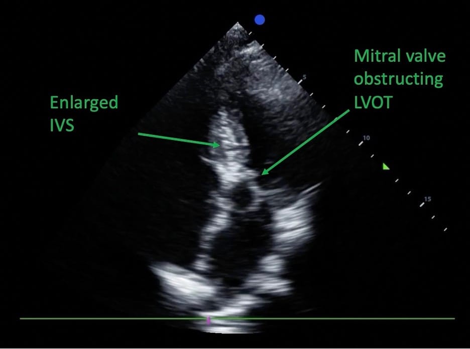
Obstructive shock may also result from dynamic left ventricular outflow tract obstruction, which in the perioperative setting could be associated with hypertrophic obstructive cardiomyopathy (HOCM).21 Typical findings of HOCM include a small, hyperdynamic LV with asymmetric septal hypertrophy, causing near-complete or partial obliteration of the ventricular cavities, and characteristic systolic anterior motion of the mitral valve, partially obstructing the LVOT during systole (see Figure 8).
Pulmonary Embolism
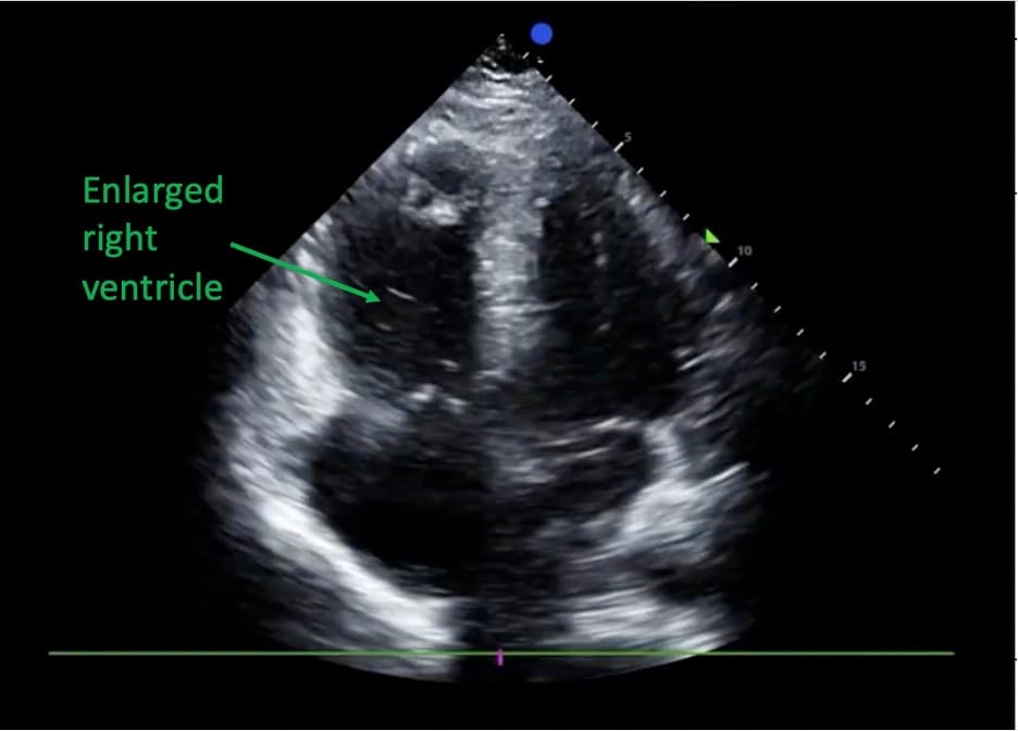
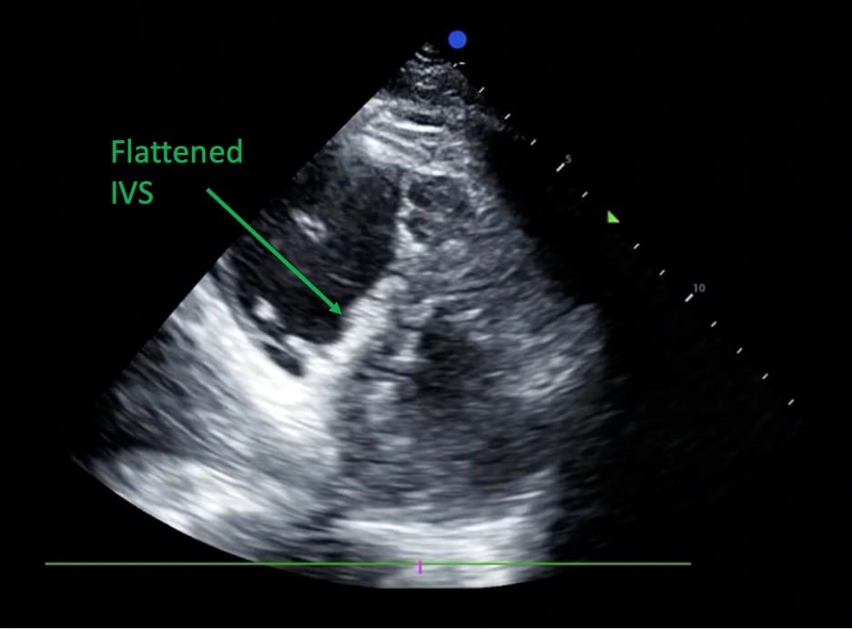
When a pulmonary embolus (PE) is present, three main useful signs may be seen on cardiac ultrasound: right ventricular dilation, septal flattening, and McConnell’s sign. RV dilatation, with the right ventricle exceeding approximately 60% of the left ventricular size, is the most common sign during PE (see Figure 9).22 The most specific echocardiographic finding associated with pulmonary embolism is the McConnell’s sign, characterized by akinesis of the right ventricular free wall with preserved apical contraction of both ventricles. Despite its high specificity, McConnell’s sign can be seen in other conditions, such as right ventricular infarction, so clinical correlation is necessary.23 Flattening of the interventricular septum or a D-shaped left ventricle is considered an indirect marker of RV strain (Figure 10).22
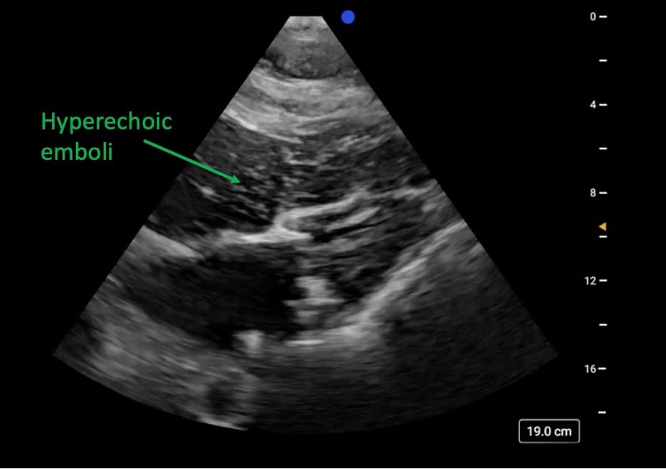
Bone Cement Implantation Syndrome (BCIS) is a perioperative pulmonary embolic phenomenon characterized by a “shower” of hyperechoic emboli during cemented orthopedic procedures such as arthroplasties.24 During cementation, the exothermic expansion of acrylic polymers elevates intramedullary pressure, driving medullary fat, marrow, bone debris, thrombi, and possibly cement into the venous circulation. This can precipitate acute pulmonary hypertension and right ventricular dysfunction, producing hypoxemia, hypotension, and in severe cases, cardiac arrest.25 Notably, a study reported high intra- and interobserver reliability in identifying embolic phenomena during hip arthroplasty using a severity rating scale based solely on the IVC view.26
Cardiogenic shock
Cardiogenic shock in the perioperative setting results from intrinsic cardiac dysfunction leading to a critical reduction in cardiac output despite adequate intravascular volume.6
Heart: When investigating cardiac function, you should assess four features with each view:
- global left ventricular systolic function
- regional wall motion abnormalities
- right vs left-sided involvement
- valvular function
If the global ventricular systolic function is reduced, you may see a left ventricle that is moderately dilated, not contracting well, and although there is some excursion of the mitral valve, it may not touch the interventricular septum (see Figure 12).6
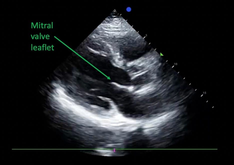
To assess regional wall motion abnormalities, the cohesive contraction of cardiac walls is often best appreciated in the PSAX view. When assessing the right versus the left heart function, the A4C and SC4C are great views for comparing all four chambers. These views, as well as the PLAX, are also good for assessing valvular function.27
IVC: The IVC may appear full.
Morrison’s Pouch and Abdomen: In Morrison’s pouch and the abdomen, you wouldn’t expect much change in the abdomen itself. However, in advanced POCUS, you may be able to assess volume congestion in the kidneys and liver.
Pulmonary: You may also see evidence of a pleural effusion or increased B lines.
The RUSH HI-MAP protocol is well-researched and has been shown to be a definitive method for quickly assessing perioperative hypotension in the emergency department setting. In this article, we amended the HI-MAP protocol to help anesthesiologists quickly discern etiologies of perioperative hypotension and shock states.





References
- Han S, Zeng T, Xie Y, et al. The effect of postoperative hypotension on surgical patients’ adverse clinical outcomes: A systematic review and meta-analysis. J Clin Anesth 2025;107:111987. https://doi.org/10.1016/j.jclinane.2025.111987
- Sessler DI, Meyhoff CS, Zimmerman NM, et al. Period-dependent associations between hypotension during and for four days after noncardiac surgery and a composite of myocardial infarction and death: a substudy of the POISE-2 trial. Anesthesiology 2018;128(2):317-27. https://doi.org/10.1097/aln.0000000000001985
- Yoshida T, Yoshida T, Noma H, et al. Diagnostic accuracy of point-of-care ultrasound for shock: a systematic review and meta-analysis. Crit Care 2023;27(1):200. https://doi.org/10.1186/s13054-023-04495-6
- Perera P, Mailhot T, Riley D, Mandavia D. The RUSH exam: Rapid Ultrasound in SHock in the evaluation of the critically lll. Emerg Med Clin North Am 2010;28(1):29-56, vii. https://doi.org/10.1016/j.emc.2009.09.010
- Cannon JW. Hemorrhagic shock. N Engl J Med 2018;378(4):370-9. https://doi.org/10.1056/NEJMra1705649
- Galderisi M, Cosyns B, Edvardsen T, et al. Standardization of adult transthoracic echocardiography reporting in agreement with recent chamber quantification, diastolic function, and heart valve disease recommendations: an expert consensus document of the European Association of Cardiovascular Imaging. Eur Heart J Cardiovasc Imaging 2017;18(12):1301-10. https://doi.org/10.1093/ehjci/jex244
- Kaptein MJ, Kaptein EM. Inferior vena cava collapsibility index: clinical validation and application for assessment of relative intravascular volume. Adv Chronic Kidney Dis 2021;28(3):218-26. https://doi.org/10.1053/j.ackd.2021.02.003
- Mistry RN, Winearls JE. Management of vasoplegic shock. BJA Educ 2025;25(2):65-73. https://doi.org/10.1016/j.bjae.2024.10.004
- Vincent JL, De Backer D. Circulatory shock. N Engl J Med 2013;369(18):1726-34. https://doi.org/10.1056/NEJMra1208943
- Vegas A, Denault A, Royse C. A bedside clinical and ultrasound-based approach to hemodynamic instability – Part II: bedside ultrasound in hemodynamic shock: continuing professional development. Can J Anaesth 2014;61(11):1008-27. https://doi.org/10.1007/s12630-014-0231-9
- Mok KL. Make it SIMPLE: enhanced shock management by focused cardiac ultrasound. J Intensive Care 2016;4:51. https://doi.org/10.1186/s40560-016-0176-x
- Miller A, Wilkinson J, Kasal J. Diagnosis of Shock States. Crit Care Clin 2025;41(3):397-428. https://doi.org/10.1016/j.ccc.2025.03.001
- Peck M WJ, Miller A. POCUS spotlight: perioperative hemodynamic assessment in noncardiac surgery. ASRA Pain Medicine News 2022;47. https://doi.org/10.52211/asra110122.048
- Kakihana Y, Ito T, Nakahara M, et al. Sepsis-induced myocardial dysfunction: pathophysiology and management. J Intensive Care 2016;4:22. https://doi.org/10.1186/s40560-016-0148-1
- Lichtenstein D. FALLS-protocol: lung ultrasound in hemodynamic assessment of shock. Heart Lung Vessel 2013;5(3):142-7.
- Volpicelli G, Elbarbary M, Blaivas M, et al. International evidence-based recommendations for point-of-care lung ultrasound. Intensive Care Med 2012;38(4):577-91. https://doi.org/10.1007/s00134-012-2513-4
- Flower L, Qawi I, Minami T. Lung ultrasound in acute respiratory failure. Crit Care Clin 2025;41(3):443-53. https://doi.org/10.1016/j.ccc.2025.02.001
- Lichtenstein D, Mezière G, Biderman P, Gepner A. The “lung point”: an ultrasound sign specific to pneumothorax. Intensive Care Med Oct 2000;26(10):1434-40. https://doi.org/10.1007/s001340000627
- Manson W HR. POCUS spotlight: lung ultrasound. ASRA Pain Medicine News 2022;47. https://doi.org/10.52211/asra020122.005
- Cardenas-Garcia J, Mayo PH. Bedside ultrasonography for the intensivist. Crit Care Clin 2015;31(1):43-66. https://doi.org/10.1016/j.ccc.2014.08.003
- Brown JM, Murtha W, Fraser J, et al. Dynamic left ventricular outflow tract obstruction in critically ill patients. Crit Care Resusc 2002;4(3):170-2.
- Kurnicka K, Lichodziejewska B, Goliszek S, et al. Echocardiographic pattern of acute pulmonary embolism: analysis of 511 consecutive patients. J Am Soc Echocardiogr 2016;29(9):907-13. https://doi.org/10.1016/j.echo.2016.05.016
- Solórzano JJC, Sánchez AM, Portillo DG, et al. Use of POCUS in shock. The other utilities of McConnell`s sign. Vis J Emerg Med 2025;39:102267. https://doi.org/10.1016/j.visj.2025.102267
- Valiton V, Giannakopoulos G, Müller H. Catastrophic bone cement implantation syndrome treated with venoarterial extracorporeal membrane oxygenation: a case report. CASE (Phila). 2022;6(1):24-6. https://doi.org/10.1016/j.case.2021.10.003
- Yang Y, Meng X, Huang Y. Study of the cement implantation syndrome: a review. Medicine (Baltimore). 2024;103(24):e38624. https://doi.org/10.1097/md.0000000000038624
- Corujo A, Irizaga G, Girón-Arango L, et al. Ultrasound monitoring to detect embolic phenomena in the inferior vena cava during hip arthroplasty. Rev Esp Anestesiol Reanim (Engl Ed). 2024;71(7):506-13. https://doi.org/10.1016/j.redare.2024.04.017
- Haskins SC, Tanaka CY, Boublik J, et al. Focused cardiac ultrasound for the regional anesthesiologist and pain specialist. Reg Anesth Pain Med 2017;42(5):632-44. https://doi.org/10.1097/aap.0000000000000650