ASRA Answers: What Is the Benefit of Using a Lateral Fluoroscopic View to Access the Interlaminar Epidural Space Compared With a Contralateral Oblique (CLO) View?
Cite as: Gill JS, Simopoulos TT. ASRA answers: what is the benefit of using a lateral fluoroscopic view to access the interlaminar epidural space compared with a contralateral oblique (CLO) view?. ASRA Pain Medicine News 2026;51. https://doi.org/10.52211/asra110126.005.
ASRA Answers
Interlaminar epidural steroid injection is one of the most common procedures performed in pain medicine. It is performed under fluoroscopic control to confirm epidural placement as well as to get the medication as close as possible to the pain-generating target.
Anteroposterior (AP) and lateral or contralateral oblique (CLO) views (the latter two are referred to as depth views) are standard for epidural access. Depth views are important to confirm epidural location because false loss of resistance is common, and a standalone AP view cannot distinguish epidural from non-epidural spread.1,2 Furthermore, direct needle trauma is an important cause of spinal cord injury, and accuracy and precision as one approaches the epidural space are paramount. Depth views provide radiologic landmarks at which the dorsal epidural space can be expected, namely the ventral interlaminar line (VILL) for the CLO view and the spinolaminar junction for the lateral view, which are critical to safety.3,4
Although the lateral view has been a standard view for epidural access, it is suboptimal in the low cervical spine and at the cervicothoracic junction because the needle tip is difficult to visualize. In two prospective comparative studies (one for cervical interlaminar access and the second for lumbar interlaminar epidural access), the CLO view was shown to be superior to the lateral view for needle tip visualization, consistency in the relationship of the needle to the VILL and the spinolaminar junction, respectively, and in plotting the cranio-caudal trajectory of the needle.3,4 In a randomized controlled trial, the CLO view was shown to be superior to the lateral view in clinically relevant parameters, including fewer needle passes and shorter needling times and a higher first attempt success rate.5 It has also been reported that routine use of the CLO view may decrease the incidence of inadvertent dural puncture during epidural access.6,7
These advantages have led to the rapid adoption of the CLO view in routine practice. A 2020 survey found that up to 65% of respondents used the CLO view at varying frequencies during epidural access for spinal cord stimulator lead placement.8
Nevertheless, the lateral view remains important to understand and use in clinical practice. The lateral view offers distinct benefits in the following scenarios.
Needle Tip Approaching Zone 2 (Usually Midline Needle) in the Cervical and Cervicothoracic Dorsal Epidural Space
A prospective study found that with a 50-degree AP obliquity, 11 out of 24 needle tips were on the VILL at loss of resistance; 20 were in Zone 1, and 4 were in Zone 2. All four Zone 2 needles were midline needles.3 The zones have been well described in a recent article on spinal nomenclature.9 (Figures 1 and 2). When a needle is beyond the VILL during cervical epidural steroid injection(CESI), it is prudent to check for epidural access with a small amount of contrast. If the needle tip approaches Zone 2, obtain an AP view to confirm it has not crossed the midline. A lateral view can further reassure you about the depth of the needle tip. This assumes that the needle tip is well visualized in the lateral view.
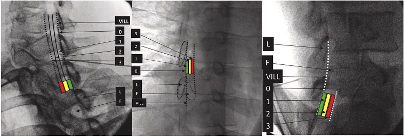
Credit: Interv Pain Med 2022;1(1). Reprinted under Creative Commons Attribution License
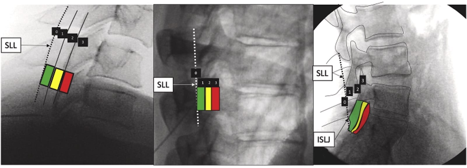
Credit: Interv Pain Med 2022;1(1) Reprinted under Creative Commons Attribution License
Inability to Use the CLO View Because of the Limitation in the Maximum Obliquity of the Fluoroscope
The recommended angle for consistent needle tip location in the CLO view is 50 degrees for cervical access and 45 degrees for lumbar access.3,4 Some fluoroscopic arms will only oblique to 30 degrees to one side. In these scenarios, one can flip the fluoroscope image intensifier below the table or have patients lie with their heads on the opposite side. Alternatively, one can accept 30 degrees of obliquity if the ventral laminar margin is visualized at the target level, and expect the loss of resistance to be anteriorly in Zone 3. These workarounds may not work well unless you are used to them, but the lateral view can still be used in this situation without issues.
Atypical Contrast Spread Patterns
A CLO view contrast spread combined with an AP view can provide a good three-dimensional understanding of contrast location. Epidural Contrast Patterns can nevertheless be myriad and difficult to interpret without other views; eg, contralateral dorsal and ipsilateral ventral spread can appear at the same depth on the contralateral oblique view, whereas on the lateral view the ventral and dorsal spread is clearly identifiable.10,11 (Figures 3 and 4). Combining the lateral view with the CLO view can provide a better understanding of contrast spread and be reassuring where the CLO spread is not reassuring.
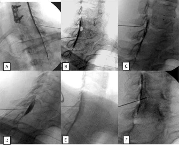
Credit: Pain Med 2017;18(6):1027-39. Reprinted with permission.
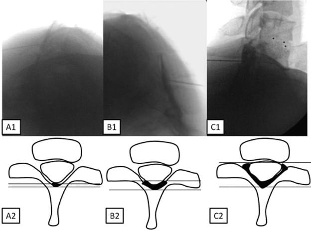
Credit: Pain Med 2017;18(6):1027-39. Reprinted with permission.
Confirming the Dorsal or Ventral location of a Catheter or a Lead
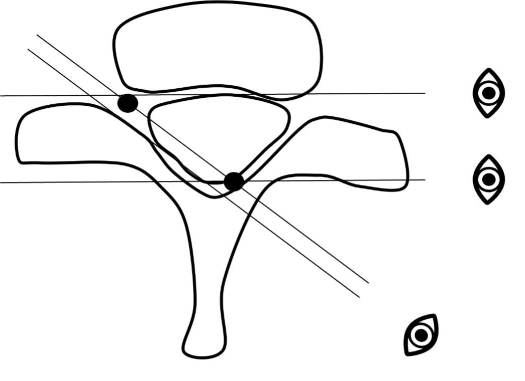
Given the CLO projection, contralateral dorsal and ipsilateral ventral objects can appear in the same plane. To truly discern and localize a catheter or lead (eg, a spinal cord stimulator lead), the lateral view is essential. (Figure 5).
Counting the Levels and Preventing Wrong-Level Errors with Transitional Anatomy
During lumbar epidural access with transitional anatomy, counting the levels is extremely important. Superimposing a lateral X-ray view with a mid-sagittal MRI provides a superior way to determine correct needle or injectate placement.
Conclusion
In conclusion, the CLO view offers distinct advantages in needle-tip visualization, trajectory planning, clear radiological landmarks, reduced risk of false loss of resistance, and improved precision. These advantages may translate to important clinical benefits, fewer needle passes, shorter needling times, a lower risk of accidental dural puncture, and, by corollary, a reduced risk of spinal cord injury—especially with cervical access.
Notwithstanding these benefits, the lateral view remains an important modality in several clinical situations:
- Deep Needle Verification: Confirming needle location when the tip extends beyond the ventral interlaminar line (VILL), especially in midline cervical placement. The lateral view can provide further reassurance about needle-tip depth, although it remains of limited utility because of difficulty visualizing the needle tip and expected variability in needle-tip location.
- Technical Constraints: Addressing equipment limitations that prevent acquiring an adequate CLO view.
- Contrast Interpretation: Evaluating and interpreting complex or atypical contrast spread patterns.
- Catheter Positioning: Confirming the precise dorsal or ventral location of an epidural catheter.
- Level Counting: Verifying anatomical levels—particularly when superimposing a midsagittal MRI over a lateral view—to ensure correct target level selection eg as in cases of transitional anatomy.


References
- Stojanovic MP, Vu TN, Caneris O, et al. The role of fluoroscopy in cervical epidural steroid injections: An analysis of contrast dispersal patterns. Spine (Phila Pa 1976) 2002;27(5):509-14. https://doi.org/10.1097/00007632-200203010-00011
- Sagir A, Simopoulos TT, Nagda JV, et al. Standalone anteroposterior contrast spread pattern is insufficient to distinguish lumbar epidural from extradural spread: A prospective study. Interv Pain Med 2025;4(3):100634. https://doi.org/10.1016/j.inpm.2025.100634
- Gill JS, Aner M, Nagda JV, et al. Contralateral oblique view is superior to lateral view for interlaminar cervical and cervicothoracic epidural access. Pain Med 2015;16(1):68-80. https://doi.org/10.1111/pme.12557
- Gill JS, Nagda JV, Aner MM, et al. Contralateral oblique view is superior to the lateral view for lumbar epidural access. Pain Med 2016;17(5):839-50. https://doi.org/10.1093/pm/pnv031
- Sim JH, Kwon HJ, Kim CS, et al. Comparison of contralateral oblique view with the lateral view for fluoroscopic-guided cervical epidural steroid injection: a randomized clinical trial. Reg Anesth Pain Med 2022;47(3):171-6. https://doi.org/10.1136/rapm-2021-103177
- Kwon HJ, Kim CS, Kim J, et al. Contralateral oblique view can prevent dural puncture in fluoroscopy-guided cervical epidural access: a prospective observational study. Reg Anesth Pain Med 2023;48(12):588-93. https://doi.org/10.1136/rapm-2022-104297
- Madan E, Hussain N, Gill JS, et al. The contralateral oblique fluoroscopic view is associated with a lower incidence of postdural puncture headache in patients undergoing percutaneous spinal cord stimulation. Pain Pract 2023;23(8):886-91. https://doi.org/10.1111/papr.13265
- Gill J, Kohan L, Hasoon J, et al. Contralateral and lateral views: analysis of the technical aspects of spinal cord stimulator lead insertion. Anesth Pain Med 2022;12(1). https://doi.org/10.5812/aapm.123357
- Gill JS, Cohen SP, Simopoulos TT, et al. A proposed nomenclature for spinal imaging and interventional procedural reporting. Interv Pain Med 2022;1(1). https://doi.org/10.1016/j.inpm.2022.100082
- Gill J, Nagda J, Aner M, et. Cervical epidural contrast spread patterns in fluoroscopic antero-posterior, lateral, and contralateral oblique view: a three-dimensional analysis. Pain Med 2017;18(6):1027-39. https://doi.org/10.1093/pm/pnw235
- Gill J, Simopoulos T, Orhurhu V, et al. Lumbar epidural contrast spread patterns for the interlaminar approach: three-dimensional analysis using antero-posterior, lateral, and contralateral oblique views. Pain Med 2020;21(4):747-56. https://doi.org/10.1093/pm/pnz256