How I Do It: A Prone Biplane Fluoroscopy-Guided Technique for Lumbar Sympathetic Nerve Block and Ablation
Cite as: Nakamura H, Hashizume K. How I do it: a prone biplane fluoroscopy-guided technique for lumbar sympathetic nerve block and ablation. ASRA Pain Medicine News 2026;51. https://doi.org/10.52211/asra110126.013.
How I Do It
Introduction
A lumbar sympathetic nerve block is a diagnostic and therapeutic procedure that targets the lumbar sympathetic trunk to interrupt sympathetic efferent activity to the lower extremities and the pelvic region.1 The procedure is indicated for conditions including complex regional pain syndrome of the lower extremities, peripheral vascular disease, lumbar shingles (herpes zoster), lumbar spine disorders, and plantar hyperhidrosis.1 Jaboulay first proposed the technique in 1924, and Felix Mandl is credited as the first to perform it using a blind approach.2 Subsequently, it has been performed primarily under fluoroscopic3 or CT guidance,4 with recent reports of ultrasound-guided approaches.5 However, there have been no formal reports to date on a lumbar sympathetic block performed in the prone position using a biplane fluoroscopy system. In this paper, we discuss our technique for performing lumbar sympathetic block in the prone position using a biplane fluoroscopy system.
Anatomy
The lumbar sympathetic chain typically consists of three to five ganglia located on the anterolateral aspect of the lumbar vertebral bodies, medial to the psoas muscle (Figure 1). The L2 and L3 ganglia are the primary clinical targets due to their abundant branching, including communicating rami.6 Fibers originating from these ganglia project to the pelvic viscera via the lumbar splanchnic nerves, and as postganglionic fibers via the communicating rami to the peripheral blood vessels, sweat glands, and nociceptors of the lower extremities, where they modulate vasomotor tone, sudomotor activity, and pain sensation.7
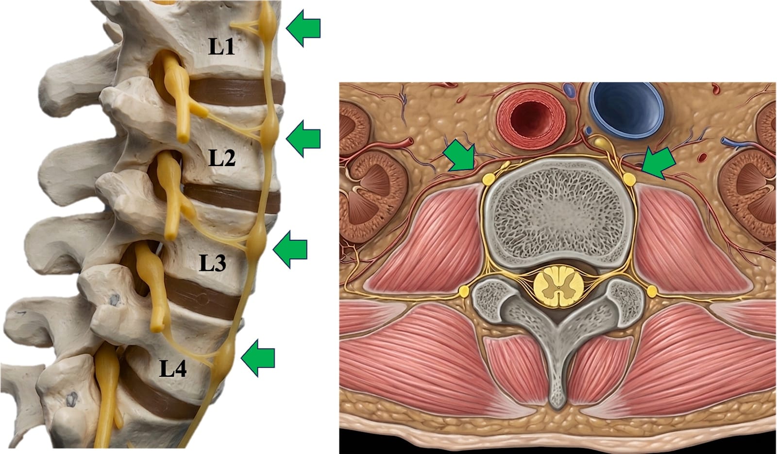
The left panel shows a lateral view of the vertebrae and sympathetic nerve, and the right panel shows a horizontal (axial) cross-section. The lumbar sympathetic chain typically consists of three to five ganglia (green arrows) located on the anterolateral aspect of the lumbar vertebral bodies, medial to the psoas muscle.
How to Match Body Position and Fluoroscopy
- Patient Positioning: Place a pillow under the patient’s abdomen to achieve flat lumbar alignment in the prone position (Figure 2).
- Fluoroscopic Alignment: Using a biplanar fluoroscopy system, establish optimal anteroposterior (AP) and lateral views.
- AP View: Optimize by positioning the spinous process centrally between the pedicles and aligning the vertebral endplates as a single line.
- Lateral View: Obtain by aligning the ribs and the posterior borders of the vertebral bodies into a single line.
- Collimation: After achieving the correct alignment, collimate (narrow the field of view) as much as possible from the beginning of the procedure.
- Fluoroscopy Mode: Use fluoroscopic low-dose pulsed mode as much as possible (our department standard is 3 frames per second). Radiation exposure increases in the lateral view because the body is thicker than in the AP view. Therefore, limit lateral fluoroscopy to single images (one-shot fluoroscopy) whenever possible, used solely to confirm needle placement and depth.
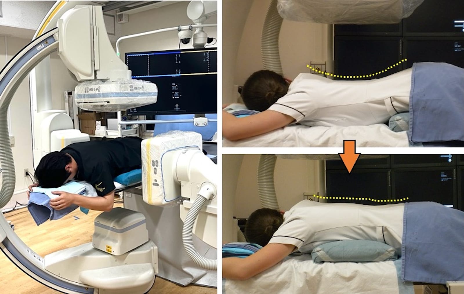
Left panel: Arrangement of the biplane fluoroscopy system.
Right panel: Patient in the prone position with a pillow placed under the abdomen to flatten the lumbar curvature (indicated by the yellow dotted line).
Points to Note and Clinical Pearls: In the early days of performing this procedure in our department, the average radiation dose per single vertebra was 10–30 milligray (mGy) for the AP tube and 30–80 mGy for the lateral tube. This has since been reduced to 5–10 mGy for the AP tube and 10–20 mGy for the lateral tube. Increased proficiency in the procedure, combined with a deliberate focus on single-image fluoroscopy—especially for lateral views—appears to reduce radiation dose.
Procedure
Equipment and Drugs: Needles: 25G or 23G 60-90 mm needles and 22G 99.5 mm needle (10 mm active tip); local anesthetic: 1% mepivacaine; contrast agent: iohexol 240 mgI/mL; block agents: approximately 2 mL of 2% lidocaine. Required disinfectants include 0.5% chlorhexidine in ethanol or povidone-iodine solution, cotton balls, gauze, Kocher forceps, and a drape.
Procedure Details: After disinfecting the procedure area with 0.5% chlorhexidine in ethanol or povidone-iodine solution, drape the area. Identify the needle insertion puncture point as follows. Measure the pedicle diameter (x cm). Mark a transverse line laterally on the side of the procedure from the inferior margin of the vertebral body. (Figure 3, Video 1). Another line is drawn at a distance of twice the diameter of the pedicle (2x cm) laterally in vertical orientation. The insertion point is at the intersection of these two lines. A local anesthetic (1% mepivacaine) is administered with a 25 G needle. Then use a 22 G 100 mm needle. Advance the needle toward the lower margin of the transverse process x cm from the target vertebral body. After slightly withdrawing the needle, advance it medially, passing beneath the transverse process. Once the needle tip is confirmed to be superior to the intervertebral foramen in the lateral view and the needle is confirmed to have contacted the mid-portion of the vertebral body, it is advanced to the anterior margin of the vertebral body using the “bevel technique” (rotating the needle to slide it along the bone surface).
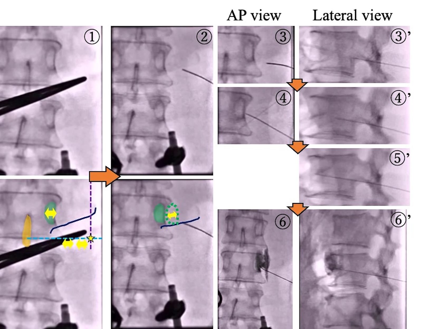
- Determine the puncture point: Measure the pedicle diameter (x cm; yellow double-headed arrow). Draw a horizontal line laterally along the inferior border of the second vertebral body (blue dotted line), and a vertical line laterally at twice the pedicle diameter (2x cm; purple dotted line). The puncture point (yellow star) is located at the intersection of these two lines. Green = pedicle, Orange = spinous process, Dark blue line = lower margin of the transverse process.
- The initial target point for the needle is at the lower margin of the transverse process, at a distance of “one pedicle width” away from the target vertebral body (indicated by the green dotted line and yellow double-headed arrow)
- Advance the needle beneath the transverse process; confirm the needle tip is superior to the intervertebral foramen on the lateral view.
- Adjust the needle position to contact the lateral aspect of the vertebral body. Confirm the needle depth and position in the lateral view.
- Advance the needle to the anterior margin of the vertebral body using the “bevel technique.”
- Confirm contrast spread along the anterior aspect of the vertebral body in the cranial-caudal direction.
Points to Note and Clinical Pearls
- If the needle contacts the lateral aspect of the vertebral body near its posterior margin, it becomes difficult to advance it to the anterior margin, even with the bevel technique. It is easier to first contact the lateral aspect of the vertebral body about 1/3 to 1/2 of the way from the anterior margin before advancing.
- If using radiofrequency, position the needle tip closer to the intervertebral disc. This is because the lumbar sympathetic ganglia tend to be located near the intervertebral discs.8
Interpretation of Contrast and Injection Of Drugs
After confirming proper placement, inject 2–3 mL of iohexol (240 mgI/mL) to verify contrast spread along the anterior aspect of the vertebral body, extending in a cranial-caudal direction. Subsequently, 1–2 mL of 2% lidocaine is injected, followed by radiofrequency thermocoagulation at 80–90°C for 120 seconds. If the patient experiences pain during the procedure, additional lidocaine is administered as needed. Measure skin temperature at the sole, regional oxygen saturation (rSO2) at the dorsum of the foot, or the perfusion index before and after coagulation.
Points to Note and Clinical Pearls
- In the AP view, a linear contrast spread extending obliquely in a caudal direction from the vertebral body (commonly referred to as a “whisker” appearance) suggests an injection into the psoas major muscle. If alcohol is injected in this setting, it will induce genitofemoral neuritis (Figure 4). If contrast spread is questionable, consider a 3D CT scan.
- If the contrast medium flows retrogradely toward the dorsal aspect of the vertebral body along the blood vessels on the lateral surface, the injected drug may reach the nerve roots or the epidural space; therefore, do not inject a neurolytic agent (Figure 4).
- In the two scenarios mentioned above, the injection of a neurolytic agent must be strictly avoided. However, in our experience, if radiofrequency is used, treatment can still be effective, and the likelihood of complications is low. The direction can often be corrected by slightly withdrawing the needle and redirecting it slightly in a cephalad or caudad direction
- This article primarily focuses on thermocoagulation for lumbar sympathetic blocks, the indications for which are described in an earlier section. However, in cases such as intractable ischemic ulcers of the toes, neurolytic lumbar sympathetic block using ethanol can be considered. When injecting alcohol, wait 5 minutes after administering 2 mL of 2% mepivacaine. Then check for sensory loss in the dermatome corresponding to the treated vertebral level, motor weakness, and sensory loss in the groin area (corresponding to the genitofemoral nerve). Next, inject ethanol at approximately 80% of the local anesthetic volume (approximately 1.6 mL). During ethanol injection, confirm under real-time fluoroscopy that the previous contrast has diluted and that no leakage occurs into other areas. If the patient experiences pain, stop the injection immediately.
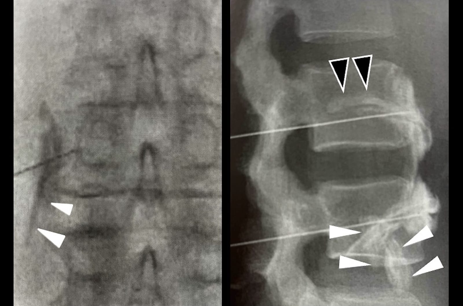
Left panel: AP view showing a linear contrast spread extending obliquely in a caudal direction from the vertebral body (commonly referred to as a “whisker” sign, white arrow).
Right panel: Contrast medium flowing retrogradely toward the dorsal aspect of the vertebral body along the blood vessels on its lateral surface (black arrow). The white arrow indicates the “whisker” sign on the lateral view.
Reprinted with permission from Morimoto M, Hashizume K, eds. Interventional Therapy for Pain Under Fluoroscopic Guidance: From Daily Clinical Procedures to the Latest Minimally Invasive Surgery. Tokyo, Japan: Kokuseido Publishing; 2020.
Complications
Potential complications include subcutaneous hemorrhage; hemorrhage or hematoma resulting from puncture of retroperitoneal blood vessels or organs; retroperitoneal abscess; and psoas abscess.
When alcohol is injected, additional complications may include genitofemoral neuritis, radicular artery embolism or spasm, spinal cord injury, and ejaculatory dysfunction. The risk of ejaculatory dysfunction is particularly relevant when ablating the L1 level in males, and caution is also required when ablating the L2 level.
Discussion
In this report, we described a technique for lumbar sympathetic nerve block performed in the prone position using a biplane fluoroscopy system.
The most widely practiced method for lumbar sympathetic nerve block is the oblique approach using a single-plane fluoroscopy system. In this technique, a needle is inserted at the lower margin of the pedicle under an oblique view, passed beneath the transverse process, and advanced to the lateral aspect of the vertebral body, where it is guided to the anterior margin using the “bevel technique” under lateral imaging.9,10 Other approaches include the transdiscal approach,11 as well as methods utilizing CT guidance12 or, more recently, ultrasound guidance.13
Based on our experience, the oblique approach often exposes the operator’s hands to radiation and may require frequent C-arm adjustments when the needle tip does not align with the intended target on the lateral view. While CT-guided procedures allow for precise identification of anatomical structures, they are associated with higher radiation doses.4,12 Ultrasound-guided methods are more accessible than fluoroscopic methods; however, visualizing deep anatomical structures can be challenging,5,13 and complications have been reported.14 Although no comparative studies exist to date, we believe that the use of a biplane system allows for the simultaneous and real-time visualization of both AP and lateral views. This facilitates easier identification of anatomical landmarks and eliminates the frequent C-arm repositioning typically required during conventional single-plane procedures.
Furthermore, as described above, this approach may reduce radiation exposure to the operator’s hands because of the distinct needle insertion technique. Additionally, we hypothesize that the biplane system involves lower radiation doses compared to CT-guided methods. Given recent reports suggesting the efficacy of lumbar sympathetic nerve block for symptom relief in cases such as failed back surgery syndrome,15 we believe this approach reduces the procedural burden on the operator when performing repeated lumbar sympathetic nerve block interventions.
As a limitation, we did not directly compare this approach with others; therefore, the specific advantages of this technique remain to be formally quantified. Further studies are warranted to compare the efficacy, procedural time, and radiation exposure of this approach with others.
Conclusions
Lumbar sympathetic nerve block in the prone position using a biplane fluoroscopy system avoids frequent C-arm adjustments and may reduce the operator’s procedural burden. Future comparative studies with other approaches are expected to further validate these findings.


References
- Dua A, Varacallo MA. Lumbar sympathetic block. In: StatPearls. Treasure Island, FL: StatPearls Publishing; 2026.
- Lee JH. Techniques and indications for sympathetic ganglion block in the lumbar spine. Int J Pain 2025;16(1):19-25. https://doi.org/10.56718/ijp.25-007
- Spiegel MA, Hingula L, Chen GH, et al. The use of L2 and L3 lumbar sympathetic blockade for cancer-related pain, an experience and recommendation in the oncologic population. Pain Med 2020;21:176-84. https://doi.org/10.1093/pm/pnz142
- Nordmann GR, Lauder GR, Grier DJ. Computed tomography guided lumbar sympathetic block for complex regional pain syndrome in a child: a case report and review. Eur J Pain 2006;10:409-12. https://doi.org/10.1016/j.ejpain.2005.05.006
- Jain D, Mohan VK, Shams A, et al. Beyond quadratus lumborum block: a technique for ultrasound guided lumbar sympathetic block. Pain Med 2022;23:2095-7. https://doi.org/10.1093/pm/pnac084
- Pontecorvo AA, Laurensio NF, Patrón TR,et al. The lumbar sympathetic trunk and its branching variability: relevance for clinical and interventional strategies. Clin Anat 2025;38:861-72. https://doi.org/10.1002/ca.24280
- Jetti R, Kadiyala B, Bolla SR. Anatomy, back, lumbar sympathetic chain. In: StatPearls. Treasure Island, FL: StatPearls Publishing; 2023.
- An JW, Koh JC, Sun JM, et al. Clinical identification of the vertebral level at which the lumbar sympathetic ganglia aggregate. Korean J Pain 2016;29:103-9. https://doi.org/10.3344/kjp.2016.29.2.103
- Morimoto M, Hashizume K, eds. Interventional Therapy for Pain Under Fluoroscopic Guidance: From Daily Clinical Procedures to the Latest Minimally Invasive Surgery. Tokyo, Japan: Kokuseido Publishing; 2020.10.
- Sakai T, Iseki M. Lumbar sympathetic nerve block. In: Ohseto K, Uchino H, Iida H, eds. Nerve Blockade and Interventional Therapy. Berlin, Germany: Springer; 2019.
- Ohno K, Oshita S. Transdiscal lumbar sympathetic block: a new technique for a chemical sympathectomy. Anesth Analg 1997;85:1312-6. https://doi.org/10.1097/00000539-199712000-00024
- Sun H, He M, Pang J, et al. Continuous lumbar sympathetic blockade enhances the effect of lumbar sympatholysis on refractory diabetic neuropathy: a randomized controlled trial. Diabetes Ther 2020;11:2647-55. https://doi.org/10.1007/s13300-020-00918-7
- Punj J, Marada S. Ultrasound lumbar sympathetic block: out of plane approach with insulated stimulation needle – case series of three patients. Indian J Anaesth 2020;64:148-50. https://doi.org/10.4103/ija.IJA_686_19
- Bai Y, Zhou X, Zeng L, et al. Diagnosis and management of a giant retroperitoneal hematoma compressing the femoral nerve, following an ultrasound-guided lumbar sympathetic block: a case report. BMC Neurol 2025;25:70. https://doi.org/10.1186/s12883-024-03808-8
- Lee JY, Sim WS, Kim J, et al. The analgesic effect of lumbar sympathetic ganglion block in patients with failed back surgery syndrome. Front Med (Lausanne). 2023;9:999655. https://doi.org/10.3389/fmed.2022.999655