How I Do It
Learn from your colleagues. ASRA Pain Medicine members share how they perform common procedures and accomplish other tasks.
- Listed by publication date, most recent on top
How I Do It: The Single-Needle Approach for Multiple Lumbar Medial Branch Blocks
Cite as: Chow RM, Stojanovic MP. How I do it: the single-needle approach for multiple lumbar medial branch blocks. ASRA Pain Medicine News 2026;51. https://doi.org/10.52211/asra110126.010.
How I Do It
Introduction
Low back pain is one of the most common and disabling complaints we see in a pain practice, and the lumbar zygapophyseal (facet) joints are a well-recognized source.1,2,3 Because patient history and physical examination cannot reliably identify the facet joint as the main pain generator, we rely on medial branch blocks (MBBs) to confirm the diagnosis before offering radiofrequency ablation.2,3,4
Each lumbar facet joint receives dual innervation from the medial branches of the dorsal rami at its corresponding level and the segment above, so a valid diagnostic block typically requires anesthetizing two medial branches per joint (and the L5 dorsal ramus for the L5–S1 joint). The conventional technique uses a separate needle for each target. In this article, we describe how we perform diagnostic lumbar MBBs at multiple contiguous levels through a single skin entry point, an approach first described by Stojanovic and colleagues.5 In our experience, this reduces the volume of local anesthetic used to anesthetize the skin, the number of skin punctures, and the number of C-arm adjustments, reducing radiation exposure to both patient and provider.
Why We Use a Single-Needle Technique
Diagnostic blocks are only useful when they are specific. Anything that anesthetizes structures other than the target medial branch raises the false-positive rate and undermines patient selection for ablation, increasing the number needed to treat.2,6,7 Potential contributors to false-positive blocks include spread of local anesthetic to the epidural space or intervertebral foramen, relief of incidental myofascial pain from excessive skin infiltration, systemic spread, and the placebo effect.2,5,8 A single-needle, single-entry approach limits skin infiltration to one site and keeps the injected volume small, which we find helps preserve specificity. It is also quicker and more comfortable for the patient, and because it uses only one C-arm position, it can reduce radiation exposure.5
Reported experience is consistent with these impressions. A prospective crossover comparison of the single- and multiple-needle techniques found comparable accuracy of final needle position and contrast spread between the two approaches.9
Anatomy
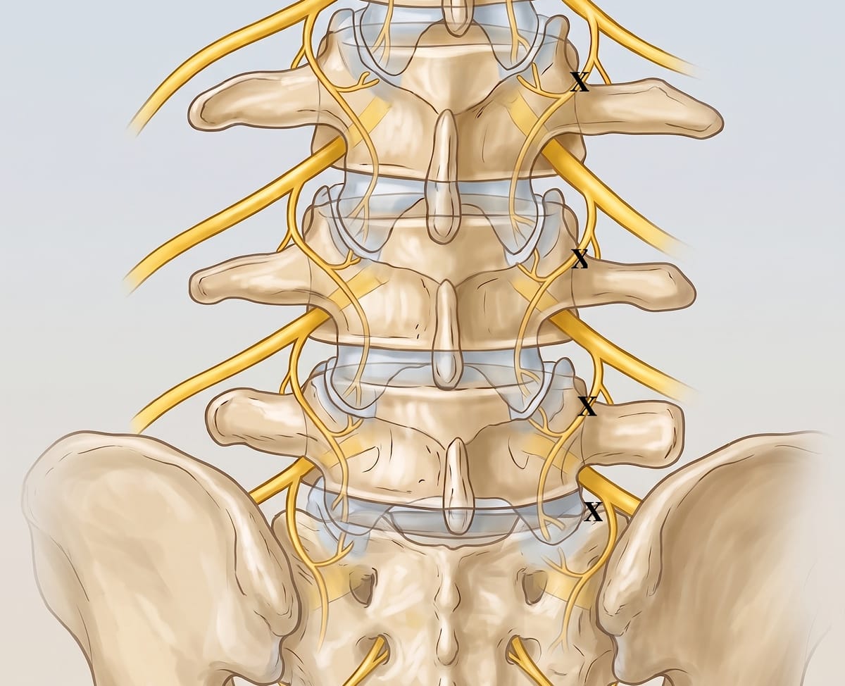
The lumbar medial branch arises from the dorsal ramus and courses across the junction of the superior articular process and the transverse process, running in the groove at the base of the superior articular process before supplying the facet joint and the multifidus.2,10 For the L1–L4 medial branches, the target is the point where the nerve crosses the dorsal surface of the transverse process (TP), just below the medial end of its superior border, near the mamillo-accessory notch or, more accurately between the base of the superior articular process (SAP) and the TP, forming a curve and the mamillo-accessory ligament.
The L5 dorsal ramus is targeted more caudally at the junction groove of the SAP of the sacrum and the superior border of the sacral ala.2,5
Indications and Patient Selection
We consider diagnostic lumbar MBBs in patients with chronic axial low back pain for whom the facet joints may be a pain generator and for whom conservative measures have not provided adequate relief.2,4,5 Patients who obtain meaningful, concordant relief from diagnostic blocks are candidates for ablation. Our threshold for a positive block uses dual comparative MBBs with ≥80% relief.2,3
The technique is most straightforward at contiguous lower lumbar levels. We find it more difficult in patients with morbid obesity, where a single anterior-posterior (AP) working view with a slight ipsilateral oblique rotation may not give reliable bony landmarks. In this scenario, we convert to a conventional multiple-needle or an oblique-view technique when needed.5 When this occurs, the use of a 22-gauge 5-inch needle is recommended.
How I Do It
Equipment and Drugs
For equipment, we utilize a 25-gauge 3.5-inch (or 22-gauge 5-inch) spinal needle, with the distal approximately 0.5 inches of the tip bent to an estimated 15–20 degrees to create a curved end in the direction of the bevel that aids in driving the needle.5,11 In terms of medication, we use non-ionic iodinated contrast and either 2% lidocaine or 0.5% bupivacaine for the MBB. We use approximately 0.1 to 0.3 mL of contrast and 0.25 mL to a maximum of 0.5 mL of local anesthetic per target with smaller volumes minimizing inadvertent spread.5,12 The skin preparation and draping are per the standard sterile technique.
The specific gauges, angles, and volumes above reflect individual practice and may vary per provider.
Positioning and Imaging
The patient is placed in a prone position, and the lower lumbar region is prepared and draped in the standard sterile fashion. Using AP fluoroscopic guidance, we see the relevant transverse processes and facet joints on the affected side.5 The procedure is then performed in the same AP view with a slight ipsilateral oblique rotation with intermittent imaging to track the needle’s movement.

Procedure
For procedures using a 25-gauge, 3.5-inch needle, local skin anesthesia is optional, as evidence suggests that creating a skin wheal can increase overall procedural discomfort.13 If a 22-gauge needle is used, a small amount of local anesthesia for the skin and subcutaneous tissues should be used.
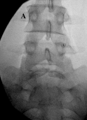
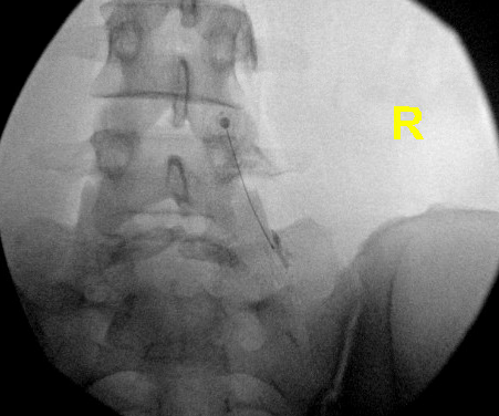
To identify a skin entry site, use the procedural 25-gauge 3.5-inch needle as a pointer to identify a fluoroscopic target. Once the target is identified, a curved needle is inserted toward the targeted medial branch of the middle level to be treated (eg L4 MB when blocking L3,4 MB and L5 DR).5 Using intermittent AP fluoroscopy and slight (5-10 degrees) ipsilateral oblique imaging, we steer the needle tip toward the first dorsal surface of the transverse process and its junction with the SAP until the needle tip contacts the bony surface and the targeted medial branch.
Once the position is satisfactory, inject 0.1-0.3 mL of contrast in increments using a tuberculin syringe to confirm that the contrast envelops the target nerve without vascular uptake and without spreading into the epidural space or intervertebral foramen. Then 0.25 mL to a maximum of 0.5 mL of the local anesthetic is injected.2,5,14
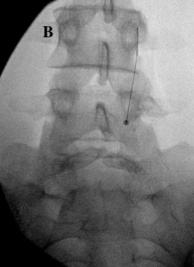
To reach the next level, withdraw the needle toward the skin without exiting, keeping the tip in the subcutaneous tissue. Then redirect the needle to the level above at a 20-30-degree angle to the skin, with the bevel facing anteriorly and medially. Once the tip crosses the inferior border of the superior-level TP, rotate the needle 180 degrees so that the bevel faces dorsally, then advance toward the target, effectively touching down at the junction between the SAP and the TP. Inject contrast, and then inject the local anesthetic.
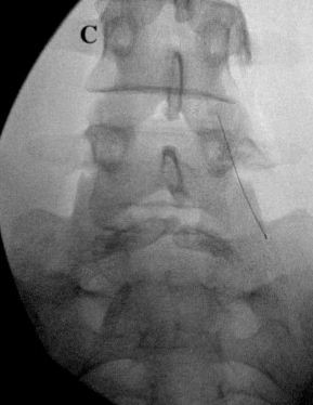
For the last level, withdraw the needle toward the skin without exiting. Then redirect the needle to the level below at a 20-30-degree angle, slightly medially when targeting another lumbar vertebral level, or at a 45-60-degree angle when targeting the sacral ala, with the bevel facing anteriorly. Once the tip crosses the top of the SAP, rotate the needle 180 degrees so that the bevel faces dorsally, and then advance to the target. Contrast is then injected, followed by the local anesthetic.5
Confirming Placement
Placement is confirmed with contrast on the AP view, potentially with slight ipsilateral oblique rotation, providing evidence of accurate placement and obviating the need for subsequent confirmatory views.5 A more extreme oblique or lateral view is available as an option when the needle tip placement or contrast spread is ambiguous.
Points to Note and Clinical Pearls
- Because a single-needle approach targets multiple branches (up to three), plan the entry site at the middle level, so the needle can be redirected appropriately.
- Keep the tip subcutaneous when repositioning, as exiting the tissue adds punctures.
- Use contrast before local anesthetic at every level, as vascular uptake or neuraxial spread should prompt repositioning before injection.14
- Keep injected volumes small to preserve diagnostic specificity.
- When using a 25-gauge 3.5-inch needle, you may not need additional local anesthetic to anesthetize the skin and subcutaneous tissues, further reducing false-positive blocks.
- Although the single-needle technique takes time to learn, it can also help with many other interventional procedures that use “out-of-plane” visuospatial needle guidance, such as sacroiliac joint injections and transforaminal epidural steroid injections.
- A 25-gauge 3.5-inch needle is preferred because it eliminates the need for superficial anesthesia and is less painful than a 22-gauge 3.5-inch needle. However, it may take slightly longer to learn the technique with a 25-gauge 3.5-inch needle.
Complications
The safety profile of this procedure follows that of multiple needle diagnostic lumbar MBBs. Potential minor issues include transient pain or bruising at the injection site and vasovagal responses. More serious but uncommon risks relate to inadvertent spread or injection. These include intravascular injection, neuraxial spread with the associated risks of medication reaching the epidural or intrathecal space, infection, and nerve injury.2,14
Discussion
The single-needle technique does not change the anatomic targets of a lumbar MBB, but it does change how we reach them. By committing to one skin entry and one fluoroscopic view, we reduce procedural pain and procedure time. With this technique, if a local anesthetic is used for skin infiltration, the volume is lower than with the multi-needle technique, reducing false-positive rates and improving the block’s diagnostic specificity.2,5 The published crossover comparison supports comparable needle-placement accuracy between the single- and multi-needle approaches.9 In addition, CT-based imaging has shown that even contrast injected a few millimeters from the ideal point can reach the target medial branch, which helps to validate this technique that relies on a single view.2
Other image-guided techniques and cadaveric studies continue to refine our understanding of needle placement and target validity for lumbar MBBs.12,15
The single-needle approach is best viewed as a practical, radiation- and time-conscious option for diagnostic blocks.
Conclusion
For diagnostic lumbar medial branch blocks at multiple contiguous levels, the single-needle, single-entry approach performed entirely in the AP view is, in our practice, an efficient, well-tolerated technique that minimizes skin punctures and injected volume while preserving accuracy.5


References
- Schwarzer AC, Wang S, Bogduk N, et al. Prevalence and clinical features of lumbar zygapophysial joint pain: a study in an Australian population with chronic low back pain. Ann Rheum Dis 1995;54:100-6. https://doi.org/10.1136/ard.54.2.100
- Boswell MV, Singh V, Staats PS, et al. Accuracy of precision diagnostic blocks in the diagnosis of chronic spinal pain of facet or zygapophysial joint origin: a systematic review. Pain Physician 2003;6:449-56.
- Falco FJE, Manchikanti L, Datta S, et al. An update of the systematic assessment of the diagnostic accuracy of lumbar facet joint nerve blocks. Pain Physician 2012;15:E869-907.
- Derby R, Melnik I, Choi J, et al. Indications for repeat diagnostic medial branch nerve blocks following a failed first medial branch nerve block. Pain Physician 2013;16:479-88.
- Stojanovic MP, Zhou Y, Hord ED, et al. Single needle approach for multiple medial branch blocks: a new technique. Clin J Pain 2003;19:134-7. https://doi.org/10.1097/00002508-200303000-00009
- Schneider BJ, Joshi M, Ehsanian R, et al. Patient-perceived duration of effect of lidocaine and bupivacaine following diagnostic medial branch blocks; a multicenter study. Interv Pain Med 2022;1:100083. https://doi.org/10.1016/j.inpm.2022.100083
- Cohen SP, Doshi TL, Constantinescu OC, et al. Effectiveness of lumbar facet joint blocks and predictive value before radiofrequency denervation: the facet treatment study (FACTS), a randomized, controlled clinical trial. Anesthesiology 2018;129(3):517-35. https://doi.org/10.1097/ALN.0000000000002274
- Cohen SP, Bhaskar A, Bhatia A, et al. Consensus practice guidelines on interventions for lumbar facet joint pain from a multispecialty, international working group. Reg Anesth Pain Med 2020;45(6):424-67. https://doi.org/10.1136/rapm-2019-101243
- Stojanovic MP, Dey D, Hord ED, et al. A prospective crossover comparison study of the single-needle and multiple-needle techniques for facet-joint medial branch block. Reg Anesth Pain Med 2005;30:484-90. https://doi.org/10.1016/j.rapm.2005.05.007
- Tran J, Chau E, Calleja M, et al. Evaluation of fluoroscopic-guided lumbar medial branch block: a cadaveric study. Interv Pain Med 2026;5:100762. https://doi.org/10.1016/j.inpm.2026.100762
- Kumar N, Agorastides ID. The curved needle technique for accessing the L5/S1 disc space. Br J Radiol 2000;73:655-7. https://doi.org/10.1259/bjr.73.870.10911790
- Wahezi SE, Alexeev E, Georgy JS, et al. Lumbar medial branch block volume-dependent dispersion patterns as a predictor for ablation success: a cadaveric study. PM R 2018;10(6):616-22. https://doi.org/10.1016/j.pmrj.2017.11.011
- Chen AS, Miccio VF, Smith CC, et al. Procedural pain during lumbar medial branch blocks with and without skin wheal anesthesia: a prospective comparative observational study. Pain Med 2019;20(4):779-83. https://doi.org/10.1093/pm/pny322
- Lee CJ, Kim YC, Shin JH, et al. Intravascular injection in lumbar medial branch block: a prospective evaluation of 1433 injections. Anesth Analg 2008;106(4):1274-8, table of contents. https://doi.org/10.1213/ane.0b013e318162c358 .
- Amrhein TJ, Joshi AB, Kranz PG, et al. Technique for CT fluoroscopy-guided lumbar medial branch blocks and radiofrequency ablation. AJR Am J Roentgenol 2016;207:1-4. https://doi.org/10.2214/AJR.15.15694