How I Do It
Learn from your colleagues. ASRA Pain Medicine members share how they perform common procedures and accomplish other tasks.
- Listed by publication date, most recent on top
How I Do It: Landmark-Based Thoracic Epidural Catheterization
Cite as: Fautsch KJ, Patel AU, Novak A, et al. How I do it: landmark-based thoracic epidural catheterization. ASRA Pain Medicine News 2026;51. https://doi.org/10.52211/asra110126.011.
How I Do It
Introduction
Thoracic epidural analgesia (TEA) is a beneficial adjunct for acute pain management during and following major abdominal, thoracic, and vascular surgeries as well as rib fractures. By continuously delivering local anesthetics and/or opioids, TEA blocks noxious stimuli at the surgical site while also establishing a dermatomal sympathectomy that attenuates the surgical stress response. Combined, these factors provide a reduction in postoperative ileus and pulmonary complications when compared to systemic opioids, establishing TEA’s role as a useful adjunct in enhanced recovery protocols.1
Although alternatives to TEA exist (ie, paravertebral, transversus abdominis plane, erector spinae plane, quadratus lumborum blockade, multimodal analgesia), few are as versatile as TEA. When optimized, TEA can provide sufficient and titratable intraoperative anesthesia as well as postoperative analgesia, covering the surgical site for days and facilitating early mobilization, faster recovery, and improved patient satisfaction.2
A landmark-based technique is used widely for TEA placement. Compared to ultrasound and fluoroscopic techniques, the landmark technique is more cost-effective, requires no additional equipment, may be quicker, and has historically been deeply embedded in training. Overall success is high in the hands of a skilled practitioner; however, its first-pass success rate may be lower than with image-guided techniques. Multiple attempts increase the risk of TEA failure, dural puncture, post-dural puncture headache, back pain, and rarely neurologic injury and epidural hematoma. While vital for successful performance, palpation of landmarks has been shown to identify the correct vertebral level only 30% of the time.3,4 Older age and obesity independently predict TEA placement failure, likely from challenging anatomy and poorly palpable landmarks.5 Primary TEA failure can result from incorrect identification of the epidural space or subsequent incorrect catheter placement, which rely heavily on tactile and visual feedback. Thus, there is an opportunity to adopt other techniques that can reduce the TEA failure rate (up to 20%).6,7 Although fluoroscopy- or ultrasound-guided techniques are increasingly being used to mitigate the risk of TEA failure, they are technically demanding.8 We predict that the landmark technique, which has been the standard of care for years, will persist due to its cost-effectiveness, speed, and ease of placement in skilled hands.9
Anatomic Considerations
Successful thoracic epidural catheter placement relies on a detailed understanding of thoracic spine anatomy and curvature. The thoracic spine consists of 12 vertebral bodies that progressively transition from cervical-like to lumbar-like from cephalad to caudad. Thoracic vertebral bodies are larger than cervical vertebral bodies, and the posterior height is slightly taller than the anterior height, resulting in a distinct kyphotic sagittal curvature.10 Spinous processes from T4-9 are aligned with a steep angulation, making a paramedian approach anatomically advantageous. (Figure 1) After T10, the thoracic vertebrae phenotypically resemble lumbar vertebrae, making a midline approach more attainable.
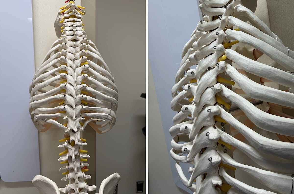
It is vital to match the dermatomal level of TEA coverage with the expected location of the surgical site. Consider placing T3-T5 for cardiovascular surgeries, T3-T7 for breast augmentation, T3-T8 for thoracotomies and chest tube placement, the mid-thoracic level (T7-9) for laparotomies, low thoracic levels (T9-10) for gynecological/urological procedures, and the level of injury for rib fractures. Confirming the dermatomal level of incision with the surgical team helps determine the optimal level of epidural placement. Identification of bony landmarks is essential; the root of the scapular spine (T3) and the inferior border of the scapula (T7) are classically used landmarks for dermatomal orientation. In patients with obesity and difficult-to-palpate anatomy, palpation of the prominent C7 spinous process can allow more consistent estimation. Alternatively, ultrasound can be used to confirm the spine level, locate the transverse process, identify the interlaminar window, and estimate epidural depth.
The epidural space surrounds the dura mater circumferentially. Nerve roots traverse the epidural space before exiting laterally through the foramen to become peripheral nerves. While accidental dural puncture during lumbar epidural placement places the needle tip near lumbar and sacral nerve roots, dural puncture during TEA placement places the needle tip near the spinal cord.11
Safety Considerations
TEA is thought to be more risky than lumbar epidural placement due to perceived increased risk of neurologic injury to the spinal cord. However, complications are fortunately rare. The most common complications include unsuccessful catheter placement and unintended dural puncture. However, serious complications, such as epidural hematomas and abscesses, permanent nerve injury, infection, and cardiovascular collapse related to local anesthetic systemic toxicity, can occur. Careful patient selection and TEA placement when the patient is relaxed, but can provide feedback, are vital. Additionally, strict aseptic technique, vigilant monitoring, and removal of the catheter when no longer needed are crucial to prevent complications. Absolute contraindications include uncorrected coagulation derangements, infection at the intended placement site, and patient refusal. Relative contraindications mirror those of neuraxial procedures, including elevated intracranial pressure, anticoagulation, thrombocytopenia, active infection, and preload-dependent states (aortic stenosis).
Positioning and Preparation
Patient Positioning and Monitoring
Before performing TEA, ensure consent has been obtained, the patient has appropriate intravenous access, monitors are in place, supplemental oxygen is available, and sedative/analgesic medications have been administered. We prefer to perform TEA while the patient is relaxed but communicative to allow feedback during the procedure. To optimize access to the interlaminar space, we recommend placing the patient in a sitting position with flexion of the cervical and thoracic spine. An epidural positioner is highly recommended. Alternatively, the lateral decubitus position can be used if the patient cannot sit for the duration of placement.
Performance of Thoracic Epidural Catheterization (Figure 2, Video 1)
Given the steep caudad angulation of thoracic spinous processes, the midline approach can be challenging. Our preferred technique is the landmark-based paramedian approach described originally by Manion et al combined with the author’s own pulsatile fluid column test to improve the success of optimal placement.12,13 The details of the steps are described below:
- After optimally positioning the patient, prepare and drape the targeted thoracic segments using strict aseptic technique. Using lidocaine, infiltrate the skin and subcutaneous tissues approximately 1 cm lateral to the inferior aspect of the targeted spinous process. Contact the ipsilateral lamina and perform local infiltration of the periosteum as well as in medial and cephalad directions to numb the intended needle paths.
- Introduce the epidural needle (Tuohy) perpendicular with the bevel directed cephalad 1 cm lateral to midline until the lamina is contacted (Figure 2A). Note the depth of first contact.
- Withdraw the needle back to the subcutaneous tissue, redirect, and advance with a slight medial angle until bony contact (Figure 2B-C). Repeat the medial revisions until the needle contacts bone (lamina) at a more superficial depth (approximately 2-5 mm) than the original depth at first perpendicular lamina contact (Figure 2D). This suggests that the needle tip is at the junction of the lamina and the spinous process. This is the intended medial angle for the rest of the technique, and no further medial angulation is necessary.
- With the same medial angle, withdraw the needle to the subcutaneous tissue and redirect the needle slightly cephalad to the same depth (Figure 2E). If you contact bone, redirect the needle slightly more cephalad and advance (Figure 2F).
- If you no longer contact bone and the depth exceeds the previous depth of bony contact, begin the loss of resistance (LOR) technique, as the needle tip may be closer to the ligamentum flavum. Many providers describe the ligamentum flavum as having a “rubbery” feel with advancement compared to the inability to advance when contacting bone.
- Remove the epidural needle stylet and attach a Luer-Lock LOR syringe filled with saline. Advance the needle slowly, millimeter by millimeter, checking for LOR between each advancement. Alternatively, you may use a continuous technique, advancing the needle and LOR syringe slowly with constant pressure on the LOR syringe.
- LOR is attained when the saline in the LOR syringe empties easily, signifying that the needle tip is in the epidural space.
- When this occurs, stabilize the epidural needle, disconnect the syringe, and thread the epidural catheter. Leave the catheter ~4-6 cm past where LOR was noted.
- Place the alligator clamp at the end of the epidural catheter; then aspirate. Aspiration should yield little to no fluid return to confirm appropriate placement. If you aspirate blood or cerebrospinal fluid (CSF), the catheter is intravascular or intrathecal, respectively, and should be removed. Additionally, we recommend using the pulsatile fluid column test to confirm appropriate epidural catheter placement (described below).
- Administer a “test dose” of a local anesthetic with epinephrine to confirm the catheter is in the epidural space. Before injection, note the patient’s heart rate. Intravascular injection may increase heart rate or cause neurologic symptoms, such as ringing in the ears. If symptoms occur, resite the epidural.
- If the patient remains asymptomatic, the test dose is considered “negative” and properly placed. Secure the catheter using a sterile occlusive dressing and tape.
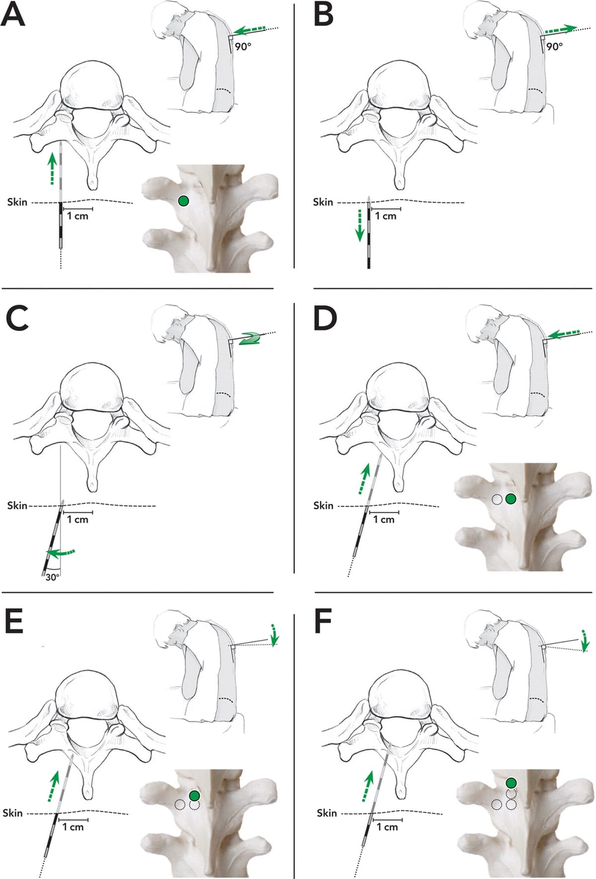
Figures 2A-F Reproduced with permission from Manion SC, Brennan TJ. Thoracic epidural analgesia and acute pain management. Anesthesiology 2011; 115(1):184.
Confirmation of Successful Placement—Pulsatile Fluid Column Test
The pulsatile fluid column test is a practical and cost-effective approach described by Selvamani et al13 to confirm correct epidural catheter tip positioning in the epidural space. This technique uses a “falling meniscus,” or the free inward movement of a fluid column within a catheter, driven by the interplay between the fluid column’s weight and the pressure difference between the epidural space and air. In turn, transmitted epidural pulsation in the falling fluid column is observed as a “pulsatile fluid column” with correct epidural positioning of the catheter tip.
After performing the epidural procedure using a standard LOR technique with saline and threading the epidural catheter to the desired length, aspirate the catheter to confirm no blood or CSF return. Then prime it with 5-8 mL of normal saline. Disconnect the alligator clamp, and elevate the end of the epidural catheter above the insertion site by about 10-20 cm. There, a fluid column is observed that drops inside the catheter. While slowly raising the outer end of the catheter, a “falling pulsatile fluid column” can be observed with the air-fluid level pausing briefly with pulsations, synchronous with the heartbeat. (Video 2) In patients with reduced epidural compliance, these pulsations can be observed at the end of a fluid-filled Tuohy needle, confirming the epidural location of the needle tip. (Video 3)
Note that this test may be difficult with a wire-reinforced or opaque catheter. In these instances, attach fluid-primed extension tubing to the epidural catheter and observe pulsatility in the larger tubing. When feasible, this simple, noninvasive bedside task can be a valuable tool for identifying catheters in the epidural space.
Troubleshooting and Clinical Pearls
Use caution to avoid a more lateral insertion point, as failure to contact the lamina or transverse process could result in needle placement in the paravertebral space or a pneumothorax from lung puncture. If the insertion point is too close to the midline, the procedure may be technically challenging due to thoracic spine angulation. Excessive medial angulation may contact spinous processes at a shallower depth than initial lamina contact, increasing procedural difficulty and limiting ability to navigate bony contact. Too much cephalad angle would result in missing the interspace at the level of entry and placing the needle at a higher interspace, noted by greater depth or even inability to reach the cephalad interspace with a standard-length Tuohy needle.
Conclusions
Providers have historically used the landmark-based technique for TEA placement because it is cost-effective, fast, and can be performed at the bedside. These factors lead to consistent, reliable placement in a busy surgical practice. However, in patients with difficult-to-palpate landmarks, because of obesity or older age, ultrasound or fluoroscopy guidance can prevent repeat attempts, false LOR, and minimize patient risk. For most patients, a landmark-based epidural technique, with the addition of the pulsatile fluid column test, allows consistent identification of the epidural space. Thus, we predict that the landmark technique, which has been the standard of care for years, will persist because of its cost-effectiveness, speed, and ease of placement in skilled hands.




References
- Pöpping DM, Elia N, Marret E, et al. Protective effects of epidural analgesia on pulmonary complications after abdominal and thoracic surgery: a meta-analysis. Arch Surg 2008;143(10):990-9. https://doi.org/10.1001/archsurg.143.10.990
- Yeung JHY, Gates S, Naidu BV, et al. Paravertebral block versus thoracic epidural for patients undergoing thoracotomy. Cochrane Database Syst Rev 2016(2). https://doi.org/10.1002/14651858.CD009121.pub2
- Perez JJ, DeFaccio RJ, Heintschel A, et al. Thoracic epidural catheter placement failure rate by contrast epidurography: a single-center retrospective study. Reg Anesth Pain Med 2026;rapm-2025-107456. https://doi.org/10.1136/rapm-2025-107456
- Tran DQ, Van Zundert TC, Aliste J, et al. Primary failure of thoracic epidural analgesia in training centers: the invisible elephant? Reg Anesth Pain Med 2016;41(3):309-13. https://doi.org/10.1097/AAP.0000000000000394
- Sutthibenjakul K, Pakpirom J, Benjhawaleemart P, et al. Real-time ultrasound-guided versus landmark techniques for thoracic epidural placement in elderly patients: A randomized comparative study. Sci Rep 2026;16(1):24868. https://doi.org/10.1038/s41598-026-55398-9
- Tran Q, Booysen K, Botha HJ. Primary failure of thoracic epidural analgesia: revisited. Reg Anesth Pain Med 2024;49(4):298-303. https://doi.org/10.1136/rapm-2023-105151
- Makino Y, Miyake K, Roche D, et al. Ultrasound guidance versus anatomical landmarks for neuraxial anaesthesia in adults. Cochrane Database Syst Rev 2025(5):CD014964. https://doi.org/10.1002/14651858.CD014964.pub2
- Pakpirom J, Thatsanapornsathit K, Kovitwanawong N, et al. Real-time ultrasound-guided versus anatomic landmark-based thoracic epidural placement: a prospective, randomized, superiority trial. BMC Anesthesiol 2022;22(1):198. https://doi.org/10.1186/s12871-022-01730-5
- Ökmen K, Yıldız DK. Landmark-guided versus real-time ultrasound-guided combined spinal-epidural anesthesia techniques: paramedian sagittal oblique and transverse interlaminar approach. J Med Ultrasound 2024;32(1):55-61. https://doi.org/10.4103/jmu.jmu_22_23
- Goh S, Price RI, Leedman PJ, et al. The relative influence of vertebral body and intervertebral disc shape on thoracic kyphosis. Clin Biomech (Bristol) 1999;14(7):439-48. https://doi.org/0.1016/s0268-0033(98)00105-3
- McLeod GA, Cumming C. Thoracic epidural anaesthesia and analgesia. Continuing Education in Anaesthesia Critical Care & Pain 2004;4(1):16-19.
- Manion SC, Brennan TJ. Thoracic epidural analgesia and acute pain management. Anesthesiology 2011;115(1):181-8. https://doi.org/10.1097/ALN.0b013e318220847c
- Selvamani BJ, Sondekoppam RV. Pulsatile fluid column—a simple bedside test to confirm optimal placement of thoracic epidural catheter. Can J Anaesth 2023;70(10):1709-10. https://doi.org/10.1007/s12630-023-02551-x