Painful Diabetic Peripheral Neuropathy: A Disorder of Metabolic Injury and Maladaptive Pain Signaling
Cite as: Sierra S, Singla P. Painful diabetic peripheral neuropathy: a disorder of metabolic injury and maladaptive pain signaling. ASRA Pain Medicine News 2026;51. https://doi.org/10.52211/asra080126.012.
The Scope of the Problem
Diabetes mellitus (DM) is a chronic metabolic disease affecting over one in nine adults worldwide, with prevalence having doubled over the past 30 years.1,2 Diabetic peripheral neuropathy (DPN) is the most common long-term complication of DM, affecting about 50% of individuals over their lifetime.3,4 It typically presents as symmetric, length-dependent distal sensory loss, autonomic impairment, and in more severe cases, motor involvement.4 Approximately one-third of patients with DPN will also develop painful diabetic neuropathy (PDPN), characterized by neuropathic pain that typically begins in the toes and gradually progresses proximally over time.4
Risk Factors
DPN and PDPN share many overlapping risk factors. (Figure 1) A meta-analysis including 16 studies indicated that duration of diabetes, older age, higher glycosylated hemoglobin A1c (HbA1c) and presence of microvascular complications are associated with significantly increased risk of DPN.5 In addition, prolonged metformin use may contribute to vitamin B12 deficiency, potentially leading to the development or worsening of DPN.6
Two recent meta-analyses evaluated risk factors for PDPN.3,7 Demographic variables that influence development of PDPN include female sex, advanced age, and geographical location. Many studies have shown consistent female predominance for development of PDPN3,7,8 suggesting sex related differences in pain processing, psychosocial factors, or hormonal influence.3,9 Advanced age has been linked to increased risk of PDPN in many studies, although this association may be confounded by diabetes duration rather than chronological age itself.3,7 Notably, geographic and ethnic variability exists, with South Asian populations showing lower overall DPN prevalence but disproportionately higher rates of painful symptoms, and studies from China and the Middle East reporting PDPN prevalence of 57%-65%, possibly reflecting gaps in screening and management.3,7 Among metabolic factors, longer diabetes duration is one of the strongest and most consistently associated risk factors for PDPN, likely reflecting cumulative inflammation, microvascular injury, and peripheral and central sensitization. In contrast, the relationship between glycemic control (HbA1c) and PDPN remains inconsistent, suggesting that non-glycemic factors, such as dyslipidemia, obesity, hypertension, and metabolic syndrome, may play substantial roles.3,7 Hypertriglyceridemia demonstrates the strongest and most consistent lipid-related association with PDPN, potentially through mechanisms involving oxidative stress and microvascular dysfunction independent of glycemic control.7 Increased waist circumference, a more reliable marker of visceral adiposity than BMI, has been associated with PDPN, along with hypertension.7 Furthermore, greater neuropathy severity is independently associated with PDPN, supporting the concept that pain is not merely a symptom but a marker of more extensive nerve injury associated with central sensitization and pain chronification. Psychological factors and sleep disorders are also important contributors. A higher burden of depressive and anxiety symptoms has been consistently associated with increased odds of PDPN.10 (Figure 1).
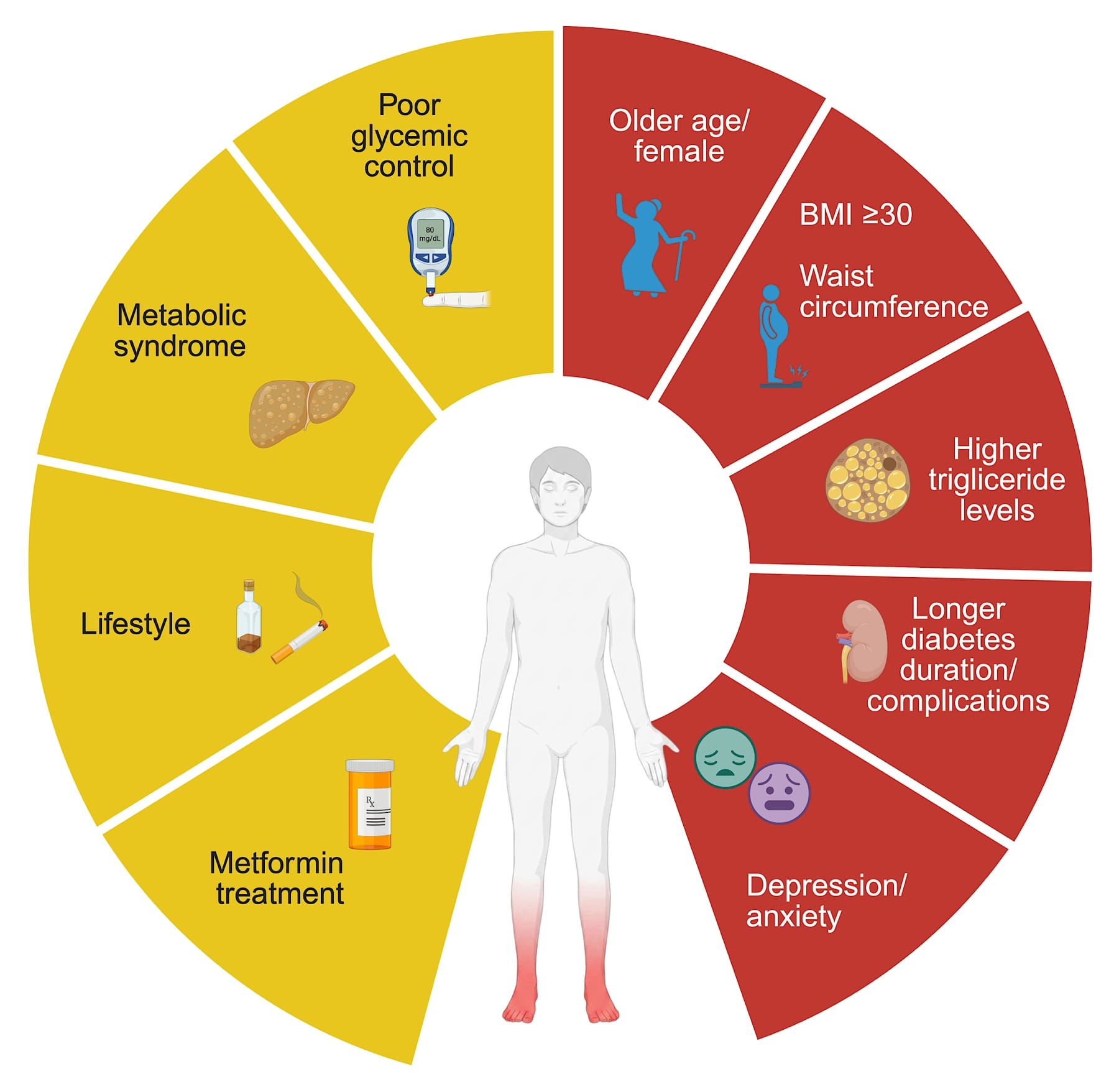
Created in BioRender. Sierra, S. (2026) https://BioRender.com/tjc0n27
Pathophysiology of Painful Diabetic Neuropathy
With regard to etiopathogenesis, the hyperglycemic state, dyslipidemia, and insulin resistance converge on oxidative stress, mitochondrial dysfunction, microvascular injury, and Schwann cell damage, affecting autonomic, large, and small nerve fibers. These metabolic derangements trigger the release of inflammatory mediators that further amplify nerve injury. Autonomic fiber degeneration manifests as abnormalities in sudomotor, cardiovascular (eg, orthostatic hypotension, resting tachycardia), gastrointestinal (eg, gastroparesis, diarrhea, or constipation), and urogenital (eg, neurogenic bladder and erectile dysfunction) function. Demyelination of large fibers (Aβ fibers) results in proprioceptive deficits and gait instability, increasing the risk of falls and foot ulcers. In contrast, degeneration of small fibers (Aδ and C fibers), together with ion channel remodeling, disrupts normal nociceptive signaling and promotes neuronal hyperexcitability.11,12
In PDPN, nociceptor sensitization and neuronal hyperexcitability arise from altered expression and post-translational modification of key ion channels in Aδ and C fiber endings. Upregulation of Nav1.8 intensifies impulse transmission in unmyelinated nociceptors, while increasing expression of transient receptor potential (TRP) channels, which sense exogenous reactive oxygen species, noxious heat, and metabolites such as methylglyoxal and hyperpolarization-activated cyclic nucleotide-gated channels (HCN2) further lower the activation threshold of peripheral nociceptors. Concurrently, downregulation of voltage-gated potassium (Kv) channels removes a critical brake on neuronal excitability, facilitating repetitive firing. These ion channel changes are accompanied by increased release of the pro-nociceptive neuropeptides substance P and calcitonin gene-related peptide (CGRP) from sensitized peripheral terminals, which amplify local neurogenic inflammation and drive ongoing nociceptive signaling toward the spinal cord. Clinically, peripheral sensitization manifests as spontaneous pain and/or exaggerated pain responses to heat (warm/hot) and mechanical (sustained pressure) stimuli at the site of injury, the hallmarks of primary hyperalgesia.
A focused history, in-office examination, and labs are paramount and usually sufficient in the diagnosis of PDN.
Growing evidence indicates that maladaptive neuroplastic changes within both the spinal cord and supraspinal structures contribute to impaired pain modulation in PDPN. At the spinal level, the sustained barrage of nociceptive input from sensitized peripheral afferents drives the release of glutamate and substance P from primary afferent central terminals into the dorsal horn. Glutamate activates postsynaptic NMDA and AMPA receptors on dorsal horn neurons, inducing long-lasting increases in excitability and increased prevalence of calcium-permeable AMPA receptors potentiates excitatory synaptic transmission. Substance P, acting via NK1 receptors, further amplifies ERK1/2 phosphorylation and NMDA receptor-mediated signaling in projection neurons. Simultaneously, spinal disinhibition occurs through reduced GABAergic, and glycinergic tonic inhibition: Downregulation of GABA-B receptors on interneurons and diminished GABA synthesis weakens the inhibitory control that normally gates nociceptive transmission, thereby unmasking and amplifying excitatory drive. Microglial activation in the dorsal horn further potentiates synaptic excitability and sustains central sensitization. At the supraspinal level, the descending pain-modulatory system, projecting from the periaqueductal gray (PAG) through the rostral ventromedial medulla (RVM) to the spinal dorsal horn, can either inhibit or facilitate nociceptive transmission.
In neuropathic pain states, the balance shifts toward facilitation: RVM ON-cells increase their activity and enhance pain signaling in the dorsal horn, while RVM OFF-cell-mediated inhibition is attenuated. Functional and structural changes in the thalamus and somatosensory cortex further promote central reorganization, amplifying pain perception beyond the degree of peripheral nerve injury.11 Although central mechanisms can amplify the pain experience in PDPN, the proportion of patients with PDPN who develop central sensitization remains poorly studied. Clinically, central sensitization manifests as the spread of sensitivity beyond the injury site in nonanatomic distributions, dynamic mechanical allodynia (pain from non-painful light-touch stimuli), and spontaneous pain. Additionally, patients may develop broader central nervous system (CNS) symptoms, including cognitive difficulties (brain fog), mood disturbances, sleep difficulties, global sensory hyperresponsiveness (heightened sensitivity to light, sound, odor, and temperature), and post-exertional malaise, reflecting the widespread nature of central nervous system dysfunction.11-13 To date, the temporal and causal relationships between peripheral and central mechanisms remain incompletely understood. It is unclear whether persistent peripheral sensitization and ectopic afferent input drive secondary CNS reorganization or whether central alterations may occur in parallel or even precede peripheral pathology. Additionally, comorbid conditions, such as metabolic syndrome, mood disorders, sleep disturbance, and systemic inflammation, may independently modulate central pain processing and influence susceptibility to PDPN (Figure 2).
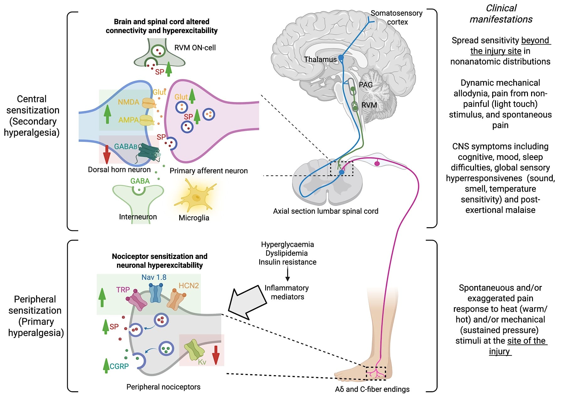
AMPA = α-amino-3-hydroxy-5-methyl-4-isoxazolepropionic acid receptor, CGRP = calcitonin gene-related peptide, CNS =central nervous system, GABA = gamma-aminobutyric acid, GABA-B = gamma-aminobutyric acid type B receptor, Glut = glutamate, HCN2 = hyperpolarization-activated cyclic nucleotide-gated channel 2, Kv = voltage-gated potassium channel, Nav1.8 = voltage-gated sodium channel 1.8, NMDA = N-methyl-D-aspartate receptor, PAG = periaqueductal gray, RVM = rostral ventromedial medulla, SP = substance P, TRP = transient receptor potential channel. Created in BioRender. Sierra, S. (2026) https://BioRender.com/oxp5hcb
Clinical Evaluation and Diagnosis
A focused history, in-office examination, and labs are paramount and usually sufficient in the diagnosis of PDN. Patients typically have a history of diabetes or prediabetes with length-dependent, and slowly (years) progressive, symmetric sensory changes beginning in the feet. On examination, assessment should include temperature and pinprick sensation to evaluate small fiber function, as well as vibration (128 Hz tuning fork), proprioception, and stance/gait to assess large fiber involvement. Laboratory evaluation should screen for common causes of neuropathy and include assessment of abnormal glucose metabolism (HbA1c, fasting glucose, or 2-hour oral glucose tolerance test), vitamin B12 level, and serum protein electrophoresis with immunofixation.14,15 Referral to neurology or electrodiagnostic testing is rarely required and is typically reserved for atypical presentations, including rapidly progressive symptoms, asymmetric sensory changes, multifocal involvement, significant weight loss, or motor-predominant weakness.12
Management of Painful Diabetic Neuropathy
Once the diagnosis is established, patients should be counseled on modifiable risk factors, including smoking cessation, limiting alcohol use, adoption of a heart‑healthy dietary pattern (eg, Mediterranean-style diet), regular exercise, and weight reduction. Optimization of cardiovascular risk factors, such as hypertension and hyperlipidemia, is also recommended. Tight glycemic control reduces the risk of developing DPN in type 2 diabetes and may modestly slow progression in type 1 diabetes.12
For PDPN, first-line therapies include tricyclic antidepressants, serotonin-norepinephrine reuptake inhibitors (SNRIs), and gabapentinoids (pregabalin or gabapentin).16 Topical agents, such as capsaicin 8% and lidocaine 5% patches, may be considered.17 (see Table 1). Although tapentadol ER is FDA-approved for PDPN, several professional societies recommend against its use because of the boxed warning for life-threatening respiratory depression, addiction, overdose, and death.
Neuromodulation remains an option for patients who have failed conservative therapies. Both high- and low-frequency spinal cord stimulation are FDA-approved for painful diabetic peripheral neuropathy, and recent meta-analyses have demonstrated greater pain relief compared with conventional medical management alone.16,18,19 (Table 1)
| Drug | Initial Dose | Maximum Dose | Side effects | Caution | NNT20 |
| TCAs(Amitryptyline/Nortriptyline) | Amitriptyline 10-25 mg qHS, Nortriptyline 10 mg qHS | Up to 100 mg | Antimuscarinic effects include dry mouth, constipation, urinary retention, and angle-closure glaucoma. Nortriptyline has fewer of these effects and is better tolerated. | Elderly patients: avoid in cardiovascular disease (QRS and QT prolongation) | 4.6 |
| SNRIs (Duloxetine) | 30 mg | 60 – 90 mg | Dry mouth, nausea, fatigue, risk of orthostatic hypotension at beginning of treatment as well as increased blood pressure, bleeding risk. and serotonin syndrome | Avoid in severe liver disease and kidney failure (eGFR <30) | 7.4 |
| Gabapentinoids | Gabapentin 100 – 300 mg qHS, Pregabalin 25 – 50 mg BID/TID | Gabapentin 1,200 – 3,600 mg divided TID, Pregabalin 300 mg divided BID | Dizziness, somnolence, peripheral edema, cognitive complaints, and gait disturbances may occur especially during the first 3-4 weeks of treatment. | Avoid in combination with opioids. Adjust dose based on kidney function. | 8.9 |
| Capsaicin 8% | Burning and erythema are common | Avoid in broken skin | 13.16 | ||
| Lidocaine 5% patch | Up to 3 patches for up to 12 hours | Mild skin reaction | Avoid in broken skin | 14.5 |
Conclusion
Painful diabetic neuropathy is a prevalent and debilitating complication of diabetes mellitus. Significant progress has been made in identifying risk factors of PDPN. Despite these advances, gaps remain, and there is a pressing need for mechanism-based treatment strategies.


References
- Genitsaridi I, Salpea P, Salim A, et al. 11th edition of the IDF Diabetes Atlas: global, regional, and national diabetes prevalence estimates for 2024 and projections for 2050. Lancet Diabetes Endocrinol 2026;14(2):149-56. https://doi.org/10.1016/S2213-8587(25)00299-2
- BD 2021 Diabetes Collaborators. Global, regional, and national burden of diabetes from 1990 to 2021, with projections of prevalence to 2050: a systematic analysis for the Global Burden of Disease Study 2021. Lancet 2023;402(10397):203-34. https://doi.org/10.1016/S0140-6736(23)01301-6
- Tao Y, Zhang HY, MacGilchrist C, et al. Prevalence and risk factors of painful diabetic neuropathy: a systematic review and meta-analysis. Diabetes Res Clin Pract 2025;222:112099. https://doi.org/10.1016/j.diabres.2025.112099
- Savelieff MG, Elafros MA, Viswanathan V, et al. The global and regional burden of diabetic peripheral neuropathy. Nat Rev Neurol 2025;21(1):17-31. https://doi.org/10.1038/s41582-024-01041-y
- Liu X, Xu Y, An M, et al. The risk factors for diabetic peripheral neuropathy: a meta-analysis. PLoS One 2019;14(2):e0212574. https://doi.org/10.1371/journal.pone.0212574
- Yang R, Yu H, Wu J, et al. Metformin treatment and risk of diabetic peripheral neuropathy in patients with type 2 diabetes mellitus in Beijing, China. Front Endocrinol (Lausanne) 2023;14:1082720. https://doi.org/10.3389/fendo.2023.1082720
- Zhou P, Zhou JS, Li JJ, et al. Prevalence and risk factors for painful diabetic peripheral neuropathy: a systematic review and meta-analysis. Front Neurol 2025;16:1564867. https://doi.org/10.3389/fneur.2025.1564867
- Elliott J, Sloan G, Stevens L, et al. Female sex is a risk factor for painful diabetic peripheral neuropathy: the EURODIAB prospective diabetes complications study. Diabetologia 2024;67(1):190-8. https://doi.org/10.1007/s00125-023-06025-z
- Mauricio D, Gratacòs M, Gómez-Peralta F, et al. The role of female sex and gender in personalised diabetes management: an overlooked perspective. Diabetes Metab Res Rev 2026;42(3):e70155. https://doi.org/10.1002/dmrr.70155
- Naranjo C, Del Reguero L, Moratalla G, et al. Anxiety, depression and sleep disorders in patients with diabetic neuropathic pain: a systematic review. Expert Rev Neurother 2019;19(12):1201-9. https://doi.org/10.1080/14737175.2019.1653760
- Zang Y, Jiang D, Zhuang X, et al. Changes in the central nervous system in diabetic neuropathy. Heliyon 2023;9(8):e18368. https://doi.org/10.1016/j.heliyon.2023.e18368
- Sloan G, Selvarajah D, Tesfaye S. Pathogenesis, diagnosis and clinical management of diabetic sensorimotor peripheral neuropathy. Nat Rev Endocrinol 2021;17(7):400-20. https://doi.org/10.1038/s41574-021-00496-z
- Fitzcharles MA, Cohen SP, Clauw DJ, et al. Nociplastic pain: towards an understanding of prevalent pain conditions. Lancet 2021;397(10289):2098-2110. https://doi.org/10.1016/S0140-6736(21)00392-5
- England JD, Gronse th GS, Franklin G, et al. Practice parameter: evaluation of distal symmetric polyneuropathy: role of laboratory and genetic testing (an evidence-based review). Report of the American Academy of Neurology, American Association of Neuromuscular and Electrodiagnostic Medicine, and American Academy of Physical Medicine and Rehabilitation. Neurology 2009;72(2):185-92. https://doi.org/10.1212/01.wnl.0000336370.51010.a1
- Mauermann ML, Staff NP. Peripheral neuropathy: a review. JAMA. 2026;335(3):255-66. https://doi.org/10.1001/jama.2025.19400
- Zhang EX, Yazdi C, Islam RK, et al. Diabetic neuropathy: a guide to pain management. Curr Pain Headache Rep 2024;28(10):1067-72. https://doi.org/10.1007/s11916-024-01293-9
- Lawson E, Singla P, Adler J, et al. Topical analgesics for neuropathic pain: an evidence-informed guide for the practicing clinician. Pain Med 2026;27(3):279-316. https://doi.org/10.1093/pm/pnaf130
- Petersen EA, Stauss TG, Scowcroft JA, et al. Effect of high-frequency (10-kHz) spinal cord stimulation in patients with painful diabetic neuropathy: a randomized clinical trial. JAMA Neurol 2021;78(6):687-98. https://doi.org/10.1001/jamaneurol.2021.0538
- Henney AE, Frank B, Riley DR, et al. Spinal cord stimulation for the treatment of painful diabetic neuropathy and risk of major adverse cardiovascular events, mortality, amputation, infection and suicide: a retrospective cohort study. EClinicalMedicine 2025;89:103489. https://doi.org/10.1016/j.eclinm.2025.103489
- Soliman N, Moisset X, Ferraro MC, et al. Pharmacotherapy and non-invasive neuromodulation for neuropathic pain: a systematic review and meta-analysis. Lancet Neurol 2025;24(5):413-28. https://doi.org/10.1016/S1474-4422(25)00068-7