How I Do It: Erector Spinae Plane Catheters for Unilateral Rib Fracture Patients
How I Do It
Cite as: Notarianni TP, Shah RH, Pozek JPJ. How I do it: erector spinae plane catheters for unilateral rib fracture patients. ASRA Pain Medicine News 2026;51. https://doi.org/10.52211/asra080126.013.
Introduction
Rib fractures occur in 15% of all traumas and 60% of patients with chest trauma.1 Rib fractures can be quite painful, especially upon deep inspiration. Splinting can result in delayed morbidity due to atelectasis, poor removal of secretions, pneumonia, and increased oxygen requirements.2 Early and adequate pain management is crucial in preventing morbidity and improving outcomes for these patients.3 The goal of rib fracture pain management is to provide sufficient analgesia while allowing the patient to actively participate in respiratory rehabilitation.4
An acute pain service (APS) consult for patients with rib fractures presenting to a level 1 trauma center was found to decrease mortality by 4.8%.5 Pain management strategies include multimodal analgesic medications and regional and neuraxial techniques, with some literature citing benefits of transcutaneous electrical nerve stimulation and cryotherapy.3 The use of neuraxial and regional anesthetic techniques may limit opioid exposure, reducing the risk of adverse sedative and respiratory depressant effects in this patient population.6 Adequate analgesia provided by these procedures under the expertise of the APS team may lead to shorter duration of intensive care unit (ICU) stay and avoidance of mechanical ventilation, which also decreases hospital and patient costs. At our institution, the current minimum daily cost of an ICU stay is approximately $10,000, with ventilator management adding $3,000 per day.
Literature Review
Since Forero et al. first described the erector spinae plane (ESP) block in 2016 as a novel regional analgesic technique for thoracic neuropathic pain, interest in its application for acute rib fracture analgesia has grown substantially.7 In 2017, the first use of a continuous ESP catheter for analgesia in a patient with four unilateral acute traumatic rib fractures was described, demonstrating substantial improvements in pain scores, respiratory mechanics, mobilization, and opioid-sparing.8 The first cohort study of 79 patients with multiple acute rib fractures who received ESP blocks was published in 2019, which reported a significant decrease in post-block maximum pain scores, a median increase of 545 mL in incentive spirometry (IS) volumes, and no change in mean arterial blood pressure from baseline as compared to pre-block values.9
Thoracic epidural analgesia (TEA) has conventionally been regarded as the gold standard regional analgesic technique for patients with multiple rib fractures because of its well-established ability to reduce pain and improve pulmonary function.10 Retrospective cohort studies comparing continuous ESP catheters with TEA have yielded mixed but generally supportive findings. In the first retrospective cohort study comparing TEA with continuous ESP block involving 52 polytrauma ICU patients with more than three unilateral posterolateral rib fractures, TEA was associated with modestly lower pain scores and reduced rescue fentanyl requirements from 12 to 48 hours after placement.11 However, early pain scores at 2, 4, and 6 hours were comparable, and both groups maintained relatively low overall pain scores throughout the study period, with the highest reported scores being 2.8 in the TEA group and 3.2 in the ESP group at 48 hours.11
Thoracic paravertebral (TPV) blocks have been regarded as an effective alternative regional technique to TEA for the treatment of acute rib fracture pain, as they offer comparable analgesia and improved respiratory function.
Kumar et al. more recently published a retrospective study involving 100 adults with unilateral traumatic rib fractures, demonstrating the non-inferiority of ESP catheters compared with TEA.12 Pain scores and total morphine consumption were similar at all evaluated time points, IS inspiratory peak volumes improved similarly in both groups, and oxygen saturation, heart rate and mean arterial pressure did not differ significantly over the observation period.12 Another 2025 retrospective study of 102 patients showed statistically lower pain scores and higher IS volumes in isolated rib fracture patients receiving continuous ESP blocks compared to those receiving TEA.13
The first randomized controlled trial comparing the two techniques was a single-blinded study of 40 adult trauma patients with three or more rib fractures, showing clinically similar morphine requirements and similarly low pain scores, but more hemodynamic stability in the ESP group, as a statistically significant drop in mean arterial blood pressures from baseline was reported in the TEA group by around 7-8 mmHg.14 In summary, the more recent literature suggests similar analgesic benefit and improvements in respiratory function in patients with rib fractures who receive ESP catheters compared with TEA, but with fewer procedural contraindications and less hemodynamic lability.11,13,14
Thoracic paravertebral (TPV) blocks have been regarded as an effective alternative regional technique to TEA for the treatment of acute rib fracture pain, as they offer comparable analgesia and improved respiratory function.10 A 2022 double-blinded randomized controlled trial of 60 patients with multiple rib fractures found similar visual analog scale pain scores at rest and during coughing, 24-hour morphine consumption, and hemodynamic parameters between groups receiving either an ESP block or a TPV block.15 A 2022 matched cohort study by Murray et al. also suggested non-inferiority of continuous ESP blocks compared to continuous TPV blocks in this patient population, describing similar analgesic benefit in terms of pain scores and time to rescue analgesics, but a greater technical ease of placing ESP catheters.16
Although TEA and TPV blocks remain effective regional techniques for rib fracture pain, their use may be limited by procedural complexity, coagulopathy, vertebral fractures, hemodynamic instability and anticoagulation administration consistent with current acute trauma care guidelines. Serratus anterior plane (SAP) blocks have emerged as another alternative because of their technical simplicity and favorable safety profile.17 While no studies have directly compared continuous ESP catheters with continuous SAP catheters, one randomized controlled trial comparing single-injection ESP and SAP blocks in patients with multiple rib fractures demonstrated superior outcomes with ESP blocks, including lower pain scores at rest and during coughing, reduced opioid requirements, and improved diaphragmatic excursion.17
Anatomical Considerations
The ESP block targets the potential space deep to the erector spinae muscle group and superficial to the transverse process of the targeted vertebral level. The transverse processes form the deep bony boundary of the injection site and serve as a consistent anatomical and sonographic landmark. A bolus injection of local anesthetic in this potential space allows extensive cranio-caudal spread along the fascial plane (Figure 1).
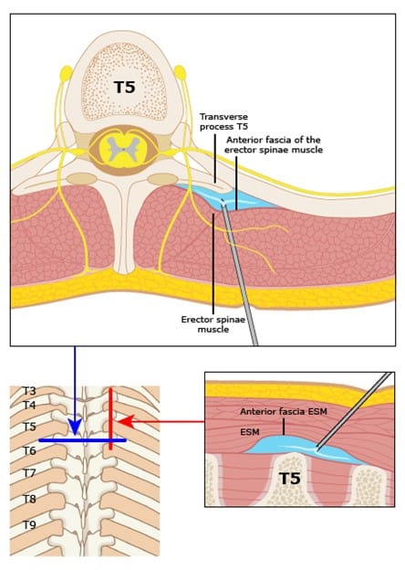
Reproduced with permission from Forero CM. Erector spinae plane block procedure guide. In: UpToDate, Connor RF (Ed), Wolters Kluwer. (Accessed on February 27, 2026.) Copyright © 2026 UpToDate, Inc. and/or its affiliates. All rights reserved.
Spinal nerves emerge from the intervertebral foramina anterior to the transverse processes and divide into dorsal and ventral rami. However, anterior paravertebral spread after standard ESP injection is inconsistent. A classically described erector spinae block injection on top of the transverse process primarily produces craniocaudal spread along the erector spinae fascial plane with consistent dorsal rami blockade, but variable and often absent anterior paravertebral spread.18,19 When the needle tip rests on the posterior surface of the transverse process tip, the injectate is deposited superficially to the superior costotransverse ligament, which acts as a barrier to anterior flow.20
A systematic review of 29 studies found that while 100% of ESP blocks spread within the erector spinae plane, only 57% reached the paravertebral space, and 51% reached the intercostal space, with highly variable extent.21 Some cadaveric studies found zero paravertebral spread with standard ESP injection.18,19
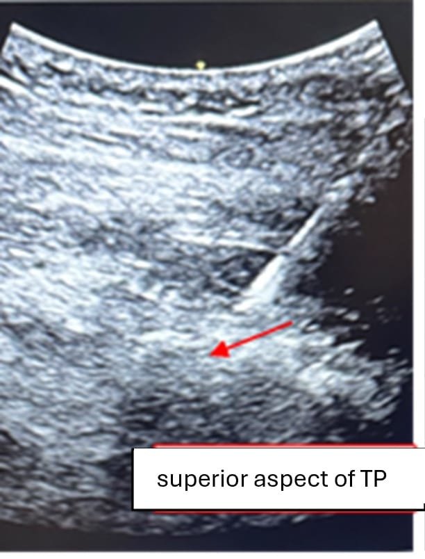
Given evidence demonstrating variable paravertebral spread following standard ESP injection, we preferentially position the needle tip near the superior aspect of the transverse process rather than directly on its posterior surface (Figure 2). We hypothesize that this trajectory may facilitate anterior migration of injectate toward the paravertebral region while maintaining the safety advantages of the ESP approach. However, the effect of this modification on injectate spread has not been definitively established.
Patient Selection
Ideal candidates for thoracic ESP catheters include patients with three or more rib fractures or complex unilateral acute rib fractures who experience significant pain inadequately controlled with standard therapy. Patients at high risk for pulmonary complications — such as older adults, those with underlying pulmonary disease, or individuals unable to effectively perform pulmonary hygiene due to pain — may derive most benefit from ESP catheter placement. It may also be advantageous in patients for whom neuraxial techniques are relatively contraindicated or technically challenging, as the target fascial plane is superficial and anatomically distant from the pleura and spinal cord. Contraindications include local infection at the insertion site, allergies to local anesthetics, transverse process fracture at the intended level, and patient refusal. Coagulopathy or the use of anticoagulants is not an absolute contraindication to ESP catheter placement; however, a pre-procedural risk-benefit analysis should be performed and thoroughly discussed with each patient during the informed consent process.
National risk stratification tools, such as the STUdy of the Management of BLunt chest wall trauma (STUMBL) score (Figure 3), can be used to guide APS consult and ICU admission. Patients with high STUMBL scores are at increased risk for pulmonary complications, including atelectasis, pneumonia, respiratory failure, prolonged hospitalization, intensive care admission, and mortality.22 These complications are largely driven by impaired pulmonary mechanics and ineffective cough secondary to severe chest wall pain, underscoring the importance of early, effective regional analgesia in high-risk rib fracture populations. At our institution, STUMBL scores of 26 or higher are admitted to the ICU. We have developed standardized criteria with the trauma team to trigger early (≤24 hours after injury) or later (>24 hours after injury) APS consultation (Table 1).
| Early Consult |
|
| Later Consult |
|
| Category | Score |
| Age | 1 point for each decade: 10–19 scores 1, 20–29 scores 2, etc. |
| Number of rib fractures | 3 points per rib fracture |
| Pre-injury anticoagulants | No = 0 Yes = 4 |
| Chronic lung disease | No = 0 Yes = 5 |
| Oxygen saturation levels | 100–95% = 0 94–90% = 2 89–85% = 4 84–80% = 6 79–75% = 8 74–70% = 10 |
(as reported by Battle et al.)
| Risk Score | Probability of Developing Complications |
| 0–10 | 13% |
| 11–15 | 29% |
| 16–20 | 52% |
| 21–25 | 70% |
| 26–30 | 80% |
| 31+ | 88% |
How I Do It:
ESP Catheter Placement
- The APS team performs the procedure with the patient in lateral decubitus or sitting position.
- After procedural time-out, infection risk is mitigated by a wide sterile prep and drape, as well as a sterile ultrasound cover.
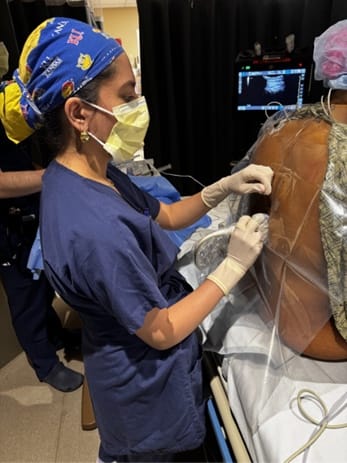
- Placing a linear or curvilinear ultrasound probe in the parasagittal plane (Figure 4), the transverse process is identified as a hyperechoic structure with an acoustic shadow, and the overlying erector spinae muscle is visualized.
- A needle is advanced in-plane toward the transverse process. Rather than depositing injectate directly on the posterior surface of the transverse process, our technique directs the needle tip deeper toward the superior aspect of the transverse process in an attempt to facilitate anterior spread toward the paravertebral space and ventral rami, while resulting predominantly in dorsal rami blockade and possible, but variable, anterior spread (Figure 2).
- Hydrodissection confirms separation of the muscle from the bony structures and creation of potential space.
- A bolus of 30mL of 0.2% ropivacaine is injected to allow cranio-caudal spread across multiple thoracic levels, and a catheter is threaded. As a fixed anatomical landmark, the transverse process guides catheter positioning and helps maintain a safe distance from the pleura, neuraxis, and major vessels.
- Following placement, the correct catheter position should be confirmed by injecting local anesthetic or a small bubble of air through the catheter under ultrasound guidance to visualize longitudinal spread deep to the erector spinae muscle.
- Catheter dislodgement and peri-catheter leakage are minimized by using adhesive anchoring devices, transparent sterile dressings, and creating a strain-relief loop.
Troubleshooting
In cases of inadequate or patchy analgesia, catheter position should be reassessed using ultrasound, with a test bolus administered to evaluate both injectate spread and clinical response. Failure to reproduce appropriate fascial plane spread or persistent inadequate analgesia despite repeat boluses should prompt consideration of catheter replacement.
Catheter Management
There is no standard regimen for achieving adequate analgesia with ESP catheters for rib fractures. While meta-analyses comparing continuous infusion (CI) and programmed intermittent boluses (PIB) for extremity and thoracic procedures exist, none compares these modalities for rib fractures.
The meta-analysis comparing modalities for thoracic procedures presents multiple different approaches, medications, doses, and regimens (Table 2). Overall, PIB significantly reduces local anesthetic consumption and provides a wider sensory blockade when compared with CI.23 A previous article from Eng et al. describes the use of PIB for achieving adequate analgesia in the rib fracture population.24
Our institution does not have PIB pumps. Instead, our APS team boluses up to 30 ml of 0.2% ropivacaine through the PNC every 12 hours, not exceeding 3 mg/kg of ideal body weight in 24 hours. This is performed bedside with appropriate periprocedural monitoring. If the patient has breakthrough pain before the next dose is due, we will consider a 10mL bolus of 0.2% ropivacaine.
The goal of the hand bolus technique is to increase the injection pressure and volume to increase the number of vertebral levels covered by the local anesthetic. Manual boluses generate significantly higher local anesthetic flow rates and pressure, which can drive bulk flow of this medication, compared with those achievable with an infusion pump at a set basal rate or PIB.25 Given the variability in number, severity, and location of rib fractures, as well as presence of chest tube, the APS team individualizes management of ESP catheters. After 4 or 5 days post-insertion, we consider the risks and benefits of continued catheter placement, with particular attention to the elevated risk of infectious complications. Daily evaluation of the PNC insertion site and dressing is recommended. Generally, ESP catheters are utilized until the patient is tolerating oral analgesics, has good respiratory function on room air, and is comfortable participating in physical therapy. If a chest tube is inserted, we routinely continue ESP catheter therapy until it is removed.
| Study | Modality/Surgery Type | PIB Settings | CI Settings |
| Taketa et al. | ESP for Thoracoscopic Surgery | Ropivacaine 0.2% 8 mL q2 hrs | Ropivacaine 0.2% 8 mL/hr |
| Eng et al. | ESP for Rib Fracture | Ropivacaine 0.2% 15 mL q3 hrs. Bolus 5 mL q1 hr. | Ropivacaine 0.2% at 8–10 mL/hr. Bolus 8 mL q1 hr. |
Conclusion
Unilateral ESP catheter as part of a multimodal analgesic plan is an effective strategy for peri-trauma pain management in patients with acute multilevel unilateral rib fractures. Management by an APS team can decrease mortality. This is an avenue for anesthesiologists to use our skills to demonstrate value to the health system, to healthcare as a whole, and most importantly, to this fragile patient population.



References
- Rogers FB, Larson NJ, Rhone A, et al. Comprehensive review of current pain management in rib fractures with practical guidelines for clinicians. J Intensive Care Med 2023;38(4):327-39. https://doi.org/10.1177/08850666221148644
- Easter A. Management of patients with multiple rib fractures. Am J Crit Care 2001;10(5):320-9.
- Bresgen TU, Salinaro F, Barcella B, et al. Acute pain management of rib fractures: a narrative review. Injury 2025;56(12):112857. https://doi.org/10.1016/j.injury.2025.112857
- Coary R, Skerritt C, Carey A, et al. New horizons in rib fracture management in the older adult. Age Ageing 2020;49(2):161-7. https://doi.org/10.1093/ageing/afz157
- Sborov KD, Dennis BM, de Oliveira Filho GR, et al. Acute pain consult and management is associated with improved mortality in rib fracture patients. Reg Anesth Pain Med Published online July 26, 2022. https://doi.org/10.1136/rapm-2022-103527
- Lin BH, Huang HM, Lin SF. Efficacy and safety of serratus anterior plane block and erector spinae plane block for rib fracture pain: a systematic review and meta-analysis. Clin J Pain 2026;42(2):e1334. https://doi.org/10.1097/AJP.0000000000001334
- Forero M, Adhikary SD, Lopez H, et al. The erector spinae plane block: a novel analgesic technique in thoracic neuropathic pain. Reg Anesth Pain Med 2016;41(5):621–7. https://doi.org/10.1097/AAP.0000000000000451
- Hamilton DL, Manickam B. Erector spinae plane block for pain relief in rib fractures. Br J Anaesth 2017;118(3):474–5. https://doi.org/10.1093/bja/aex013
- Adhikary SD, Liu WM, Fuller E, et al. The effect of erector spinae plane block on respiratory and analgesic outcomes in multiple rib fractures: a retrospective cohort study. Anaesthesia 2019;74(5):585–93. https://doi.org/10.1111/anae.14579
- van Zyl T, Ho AMH, Klar G, et al. Analgesia for rib fractures: a narrative review. Can J Anaesth 2024;71:535-47. https://doi.org/10.1007/s12630-024-02725-1
- Diwan SM, Adhye B, Nair A, et al. Comparison of thoracic epidural and thoracic erector spinae plane block for pain relief of posterolateral rib fractures: a retrospective cohort study. Ain-Shams J Anesthesiol 2022;14:87.
- Kumar M, Singh RB, Kumar A, et al. A comparative study of erector spinae plane block and thoracic epidural block on respiratory, analgesic, and hemodynamic outcomes in patients with traumatic rib fractures. Cureus 2025;17(5):e84309. https://doi.org/10.7759/cureus.84309
- Khurshid MH, Hoffman JP, Al Ma’ani M, et al. Who hits the target? erector spinae plane block versus epidural analgesia in traumatic rib fractures. J Surg Res 2025;315:923-9. https://doi.org/10.1016/j.jss.2025.10.018
- Singh S et al. Comparison of safety and efficacy of thoracic epidural block and erector spinae plane block for analgesia in patients with multiple rib fractures: a pilot single-blinded, randomised controlled trial. Indian J Anaesth 2023;67:614-9. https://doi.org/10.4103/ija.ija_844_21
- Elawamy A, Morsy MR, Ahmed MAY. Comparison of thoracic erector spinae plane block with thoracic paravertebral block for pain management in patients with unilateral multiple fractured ribs. Pain Physician 2022;25(6):483-90.
- Murray N, Swierczek J, Riley B, et al. Erector spinae plane versus paravertebral catheter techniques for rib fracture analgesia: a pilot matched cohort study. Anaesth Intensive Care 2023;51(4):348-52. https://doi.org/10.1177/14604086221106849
- El Malla DA, Helal RAEF, Zidan TAM, et al. The effect of erector spinae block versus serratus plane block on pain scores and diaphragmatic excursion in multiple rib fractures. a prospective randomized trial. Pain Med. 2022;23(3):448-55. https://doi.org/10.1093/pm/pnab214
- Luchsinger M, Varela V, Diwan S, et al. Erector spinae plane infiltration and anterior rami of spinal nerve: a cadaveric study. Reg Anesth Pain Med 2025;50(10):815-9. https://doi.org/10.1136/rapm-2024-105691
- Aponte A, Sala-Blanch X, Prats-Galino A, et al. Anatomical evaluation of the extent of spread in the erector spinae plane block: a cadaveric study. Can J Anaesth 2019;66(8):886-93. https://doi.org/10.1007/s12630-019-01399-4
- Zhang YC, Sun Y, Li SH, et al. Clinical effects, mechanisms and spread of erector spinae plane block and paravertebral block in thoracic and breast surgery: a narrative review. Int J Surg 2025;111(12):9507-19. https://doi.org/10.1097/JS9.0000000000003135
- Sun Q, Zhang C, Liu S, et al. Efficacy of erector spinae plane block for postoperative analgesia in lumbar surgery: a systematic review and meta-analysis. BMC Anesthesiology 2023;23:54. https://doi.org/10.1186/s12871-023-02013-3
- Battle CE, Hutchings H, James K, et al. The risk factors for the development of complications during the recovery phase following blunt chest wall trauma: a retrospective study. Injury 2013;44(9):1171-6. https://doi.org/10.1016/j.injury.2012.05.019
- Ren D, Liu Z, Gao Y, et al. Programmed intermittent bolus versus continuous infusion for regional analgesia following thoracoscopic surgery: a systematic review and meta-analysis. J Pain Res 2025;18:4073-83. https://doi.org/10.2147/JPR.S530018
- Eng HC, Chin KJ, Adhikary SD. How I do it: erector spinae block for rib fractures: The Penn State health experience. ASRA Pain Medicine News 2020;45
- Younger JD, Faryami A, Prasad M, et al. Direct comparison of peak bulk flow rate of programmable intermittent epidural bolus and manual epidural bolus using a closed-end multiorifice catheter: an experimental study. Anesth Analg 2023;136(6):1198-1205. https://doi.org/10.1213/ANE.0000000000006268