Diagnostic Ultrasound: The Basics of Shoulder Ultrasound
Cite as: Mauriello JP, Patel D, Barbari C, et al. Diagnostic ultrasound: the basics. ASRA Pain Medicine News 2026;51. https://doi.org/10.52211/asra080126.010.
The Shoulder
Diagnostic musculoskeletal ultrasound is being increasingly used as a diagnostic imaging modality. The goal of this newsletter series is to introduce the basics of diagnostic musculoskeletal ultrasound, starting with high-yield joints, such as the knee, shoulder, and hip. The first article in this series focused on the knee joint. In this article, we discuss the shoulder joint and review frequently encountered pathology as well as practical, image-guided “how-to” content designed for learners and clinicians at all levels.
Introduction
Ultrasound is a non-invasive imaging modality that uses high-frequency sound waves to visualize soft tissues in real time. It is widely used across medical specialties due to its safety, portability, and cost-effectiveness. Diagnostic ultrasound, particularly in musculoskeletal medicine, allows for dynamic assessment of tendons, ligaments, bursae, and joint spaces, making it an ideal tool for evaluating shoulder pain and guiding clinical decision-making at the bedside.
Basic Considerations:
A diagnostic shoulder ultrasound can be performed with the patient in a supine or seated position, depending on the region of interest and patient comfort. In ultrasound imaging, each structure should be scanned in both longitudinal and transverse planes. Anisotropy, an artifact common in tendon imaging in which structures appear artificially hypoechoic due to beam angle or insufficient gel, must be corrected to avoid misdiagnosis.1 Dynamic scanning enhances functional assessment, allowing the examiner to evaluate joint effusions during compression, tendon movement and impingement during range of motion, and ligament integrity with stress maneuvers.2
Transducer/Positioning:
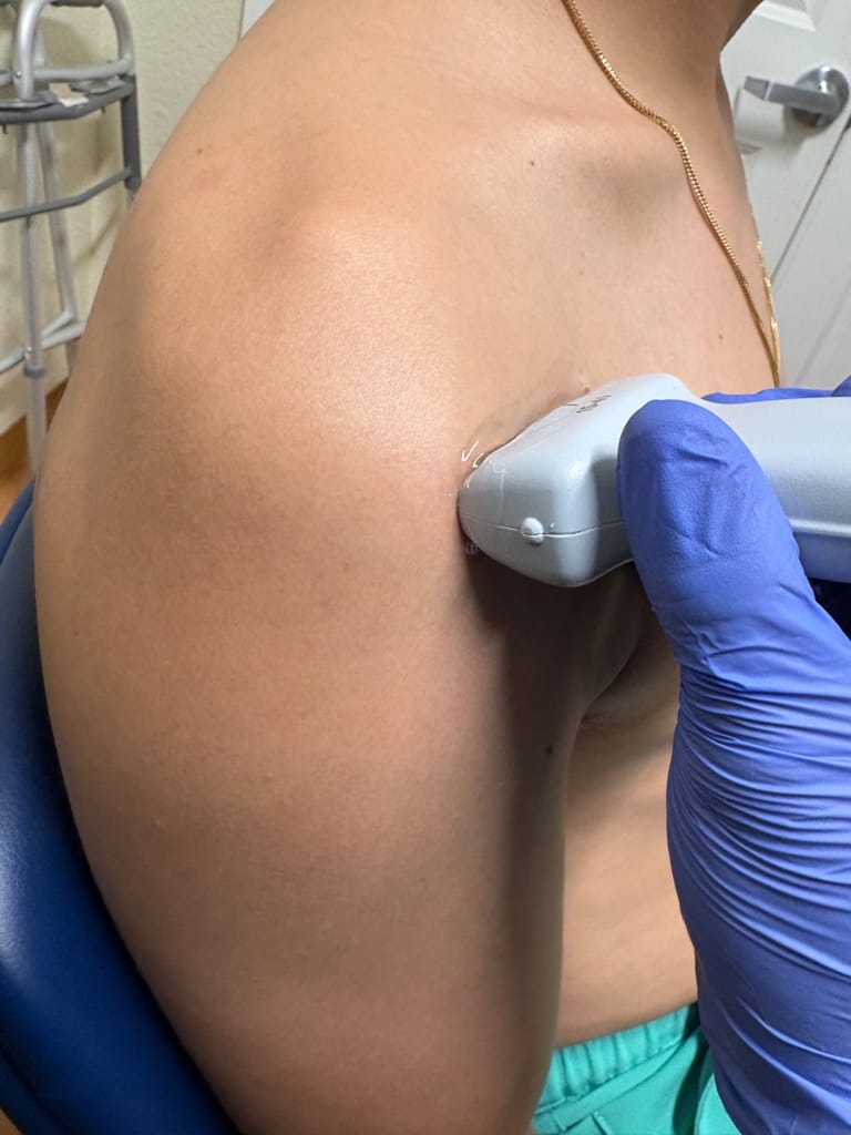
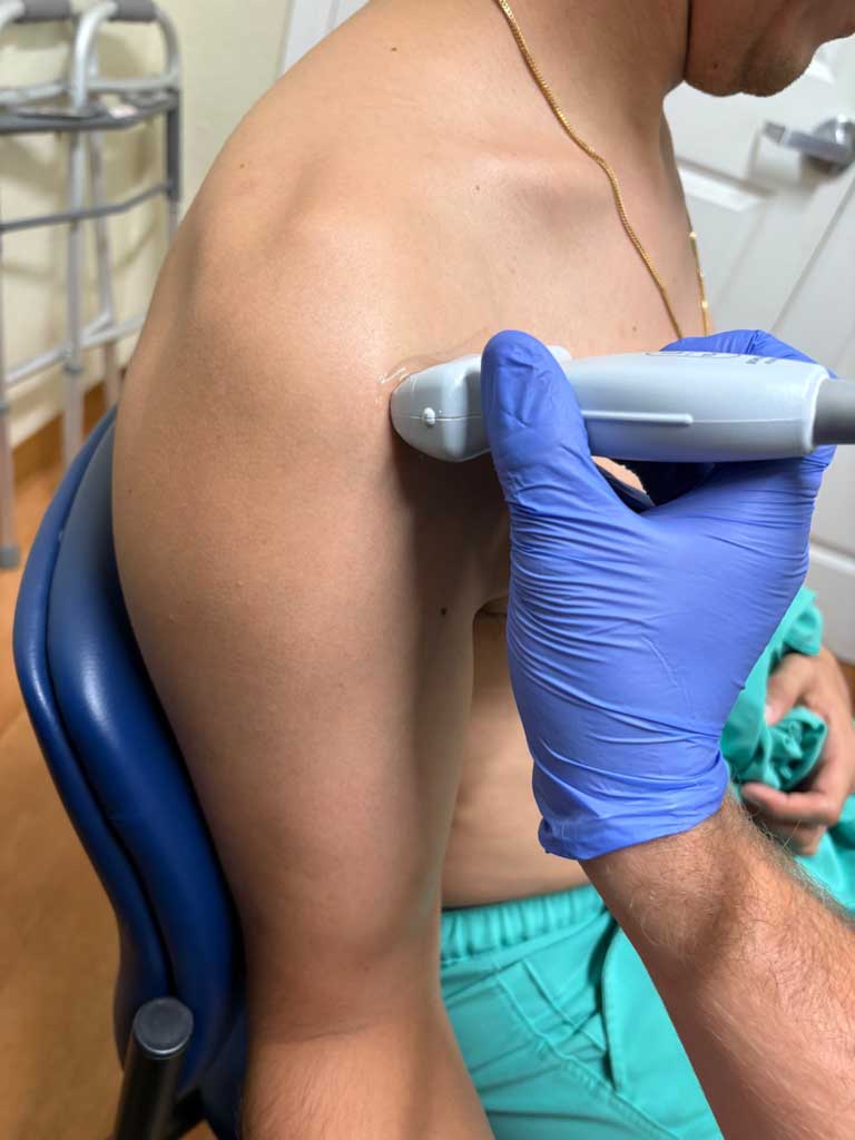
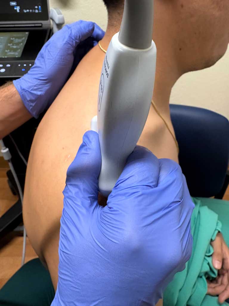
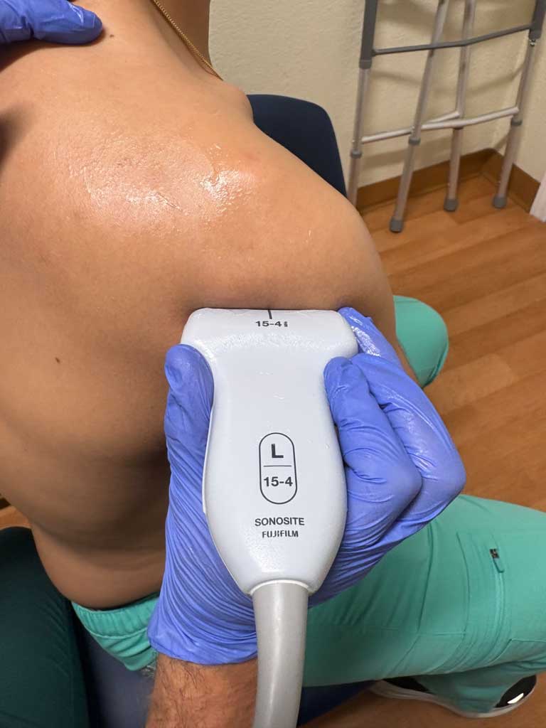
A high-frequency linear transducer is recommended. Switch to lower frequency for larger body habitus or deeper structures. For the anterior shoulder, visualization is best obtained with the patient seated, elbow flexed to 90 degrees (refer to Figures 1A–C). For the posterior shoulder, visualization is best noted when the patient is seated with the shoulder slightly adducted or crossing the arm to grab the opposite shoulder (Figure 1D).
Anterior Shoulder
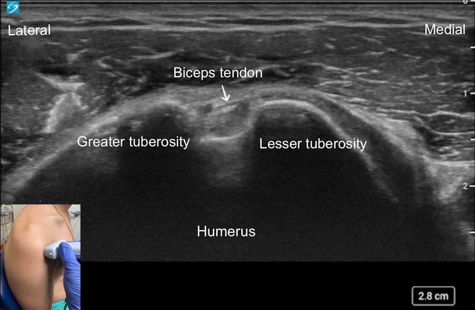
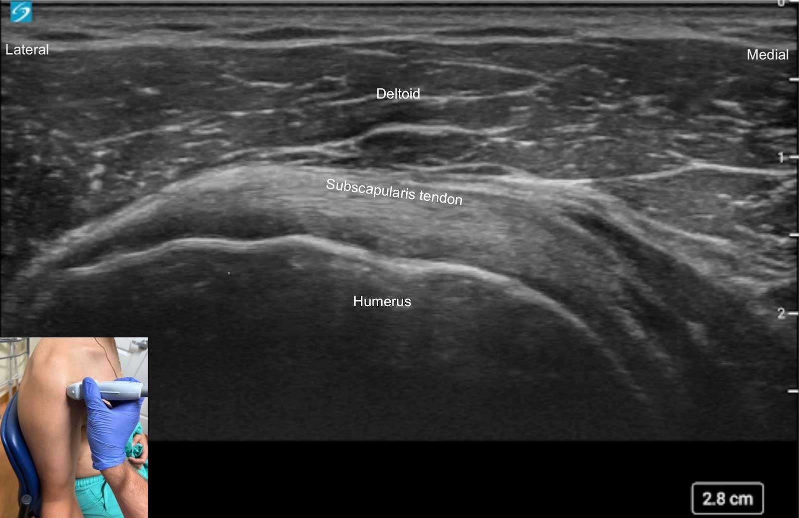
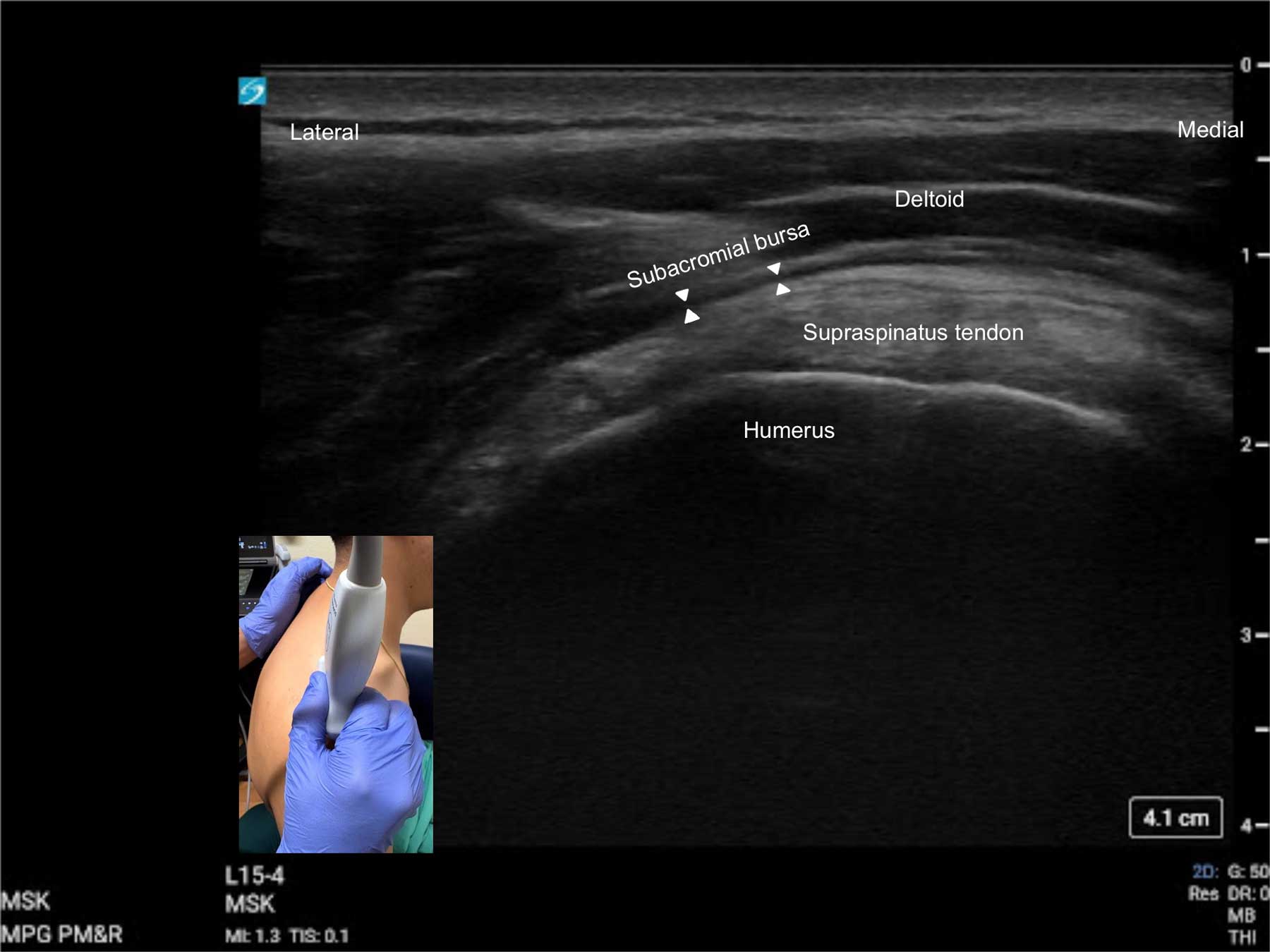
Begin by placing the transducer in short axis over the long head of the biceps tendon within the bicipital groove (Figures 1A and 3A). Scan proximally and distally to assess the extent and severity of any tears or tendinopathy as well as for any signs of fluid. Subluxation of the biceps tendon can also be assessed in this view with dynamic evaluation by externally and internally rotating the shoulder. While keeping the probe in the same orientation, externally rotate the arm and scan medially from the “home base” to view the subscapularis tendon and muscle in long axis (Figures 1B and 3B). Returning the patient’s arm to neutral position, the supraspinatus tendon can be evaluated by placing the transducer in a long axis orientation just anterior to the acromioclavicular joint (Figures 1C and 3C). The tendon lies just above the humerus. Note any anechoic defects within the tendon; scanning slightly lateral along the tendon to better visualize insertional tears, which may reveal cortical irregularities. Between the deeper supraspinatus tendon and the more superficial deltoid muscle lies the thin subacromial bursa, a potential space for fluid. Dynamic movement of the shoulder at this position can help assess for impingement. Note: The above may also be done in the crass or modified crass position.
Key Structures:
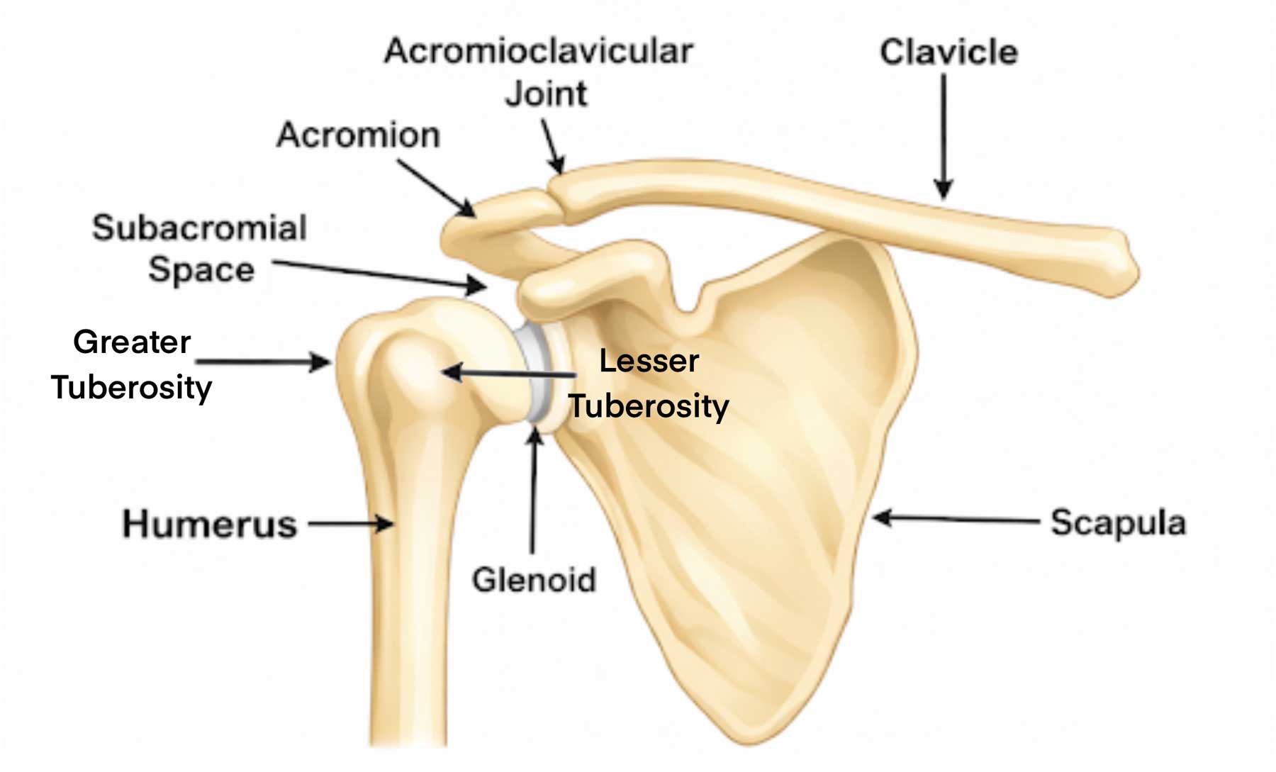
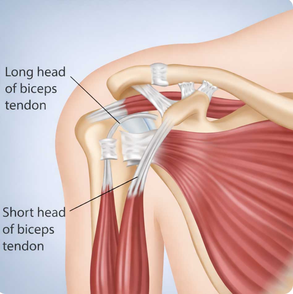
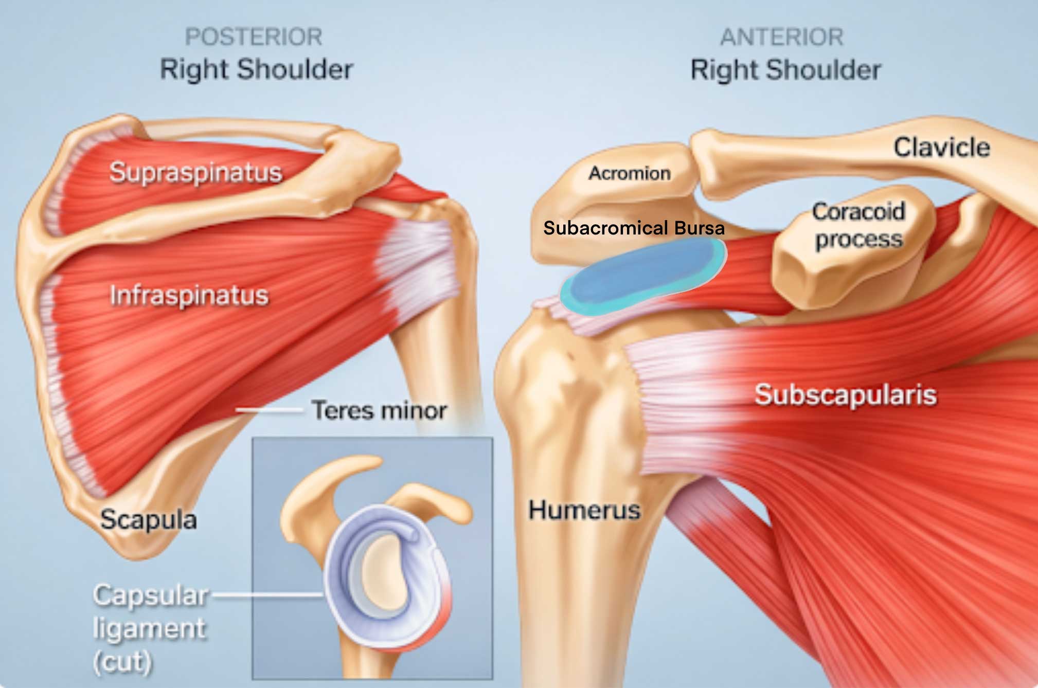
Humerus, bicipital groove (between the greater and lesser trochanters), tendon of the long head of the biceps, subscapularis, deltoid, supraspinatus, and subacromial bursa (also known as the subdeltoid bursa) (Figures 2A–C).
Pathology:
Tendinopathies are thickened, hypoechoic tendons +/- calcifications and Doppler hyperemia; There may be anechoic/hypoechoic fluid surrounding the tendon sheath with tenosynovitis.
Tendon tears are hypoechoic disruptions of tendon fibers; cortical irregularities at the tendon attachment points indicate an insertional tear.
Bursitis is anechoic/hypoechoic fluid with bursal expansion; Doppler/irregular lining suggests synovitis.
Subluxation is tendon translation out of a confined area with dynamic studies, such as with the biceps tendon.
Posterior Shoulder
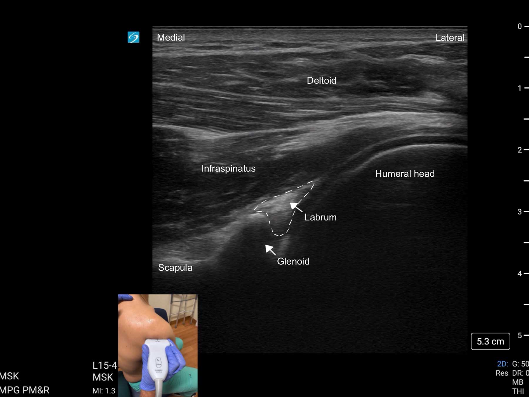
Begin by placing the transducer longitudinally to the spine of the scapula. Sweep the probe just inferior to the spine of the scapula to visualize the infraspinatus in the long axis. While maintaining the same orientation, slide the probe laterally along the infraspinatus fibers until the infraspinatus, glenoid, and humerus are visualized. Sweep the probe superior or inferior or fan until the infraspinatus, deltoid, and glenohumeral joint can be visualized in one view. The glenoid labrum may also be seen from this view (Figures 1D and 4).
Key Structures:
Infraspinatus, deltoid, glenohumeral joint, and shoulder labrum (Figure 2C)
Pathology:
Tendinopathies are thickened, hypoechoic tendons +/- calcifications and Doppler hyperemia; there may be anechoic/hypoechoic fluid surrounding the tendon sheath with tenosynovitis.
Tendon tears are hypoechoic disruptions of tendon fibers; cortical irregularities at the tendon attachment points indicate an insertional tear.
A labral tear is a hypoechoic cleft within the labrum or the complete absence of the normal hyperechoic triangular architecture of the labrum
Treatment and Next Steps
Tendinopathy: Activity modification, eccentric rehab (eg rotator cuff muscle strengthening), and/or guided injection/needling.
Bursitis: Activity modification (eg limiting overhead lifting for subacromial bursitis), topical or oral NSAIDs, physical therapy, and/or guided injection.
Labral tear: MRI arthrogram
Normal scan plus persistent mechanical symptoms: Physical therapy (if high suspicion for frozen shoulder) or MRI if there is concern for intraarticular pathology.
Summary
Diagnostic ultrasound is a valuable, real-time imaging tool that enhances the evaluation of shoulder pain by providing detailed views of soft tissue structures and joint pathology. Its accessibility, safety, and dynamic capabilities make it an essential part of musculoskeletal assessment and a practical guide for both diagnosis and treatment planning. As technology advances, its role in clinical decision-making continues to grow.
When diagnosing full-thickness rotator cuff tears, ultrasound has been shown to be extremely accurate with a sensitivity of 92% and specificity of 93%, comparable to MRI at 94%. However, it should be noted that the sensitivity of ultrasound for partial-thickness tears does decrease substantially (52%), and results should be interpreted with caution.3 Ultrasound may provide a relatively inexpensive and portable option for clinicians in the office setting. Additionally, it may be a first line option for those patients with claustrophobia and positioning constraints or those with metallic hardware or other MRI-incompatible devices.4




References
- Wu WT, Chang KV, Hsu YC, et al.. Artifacts in musculoskeletal ultrasonography: from physics to clinics. Diagnostics (Basel) 2020;10(9):645. https://doi.org/10.3390/diagnostics10090645
- Serpi F, Albano D, Rapisarda S, et al. Shoulder ultrasound: current concepts and future perspectives. J Ultrason 2021;21(85):e154-e61. https://doi.org/10.15557/JoU.2021.0025
- Lenza M, Buchbinder R, Takwoingi Y, et al. Magnetic resonance imaging, magnetic resonance arthrography and ultrasonography for assessing rotator cuff tears in people with shoulder pain for whom surgery is being considered. Cochrane Database Syst Rev 2013;2013(9): CD009020. https://doi.org/10.1002/14651858 .
- Laur O, Ha AS, Bartolotta RJ, et al. Expert panel on musculoskeletal imaging. ACR Appropriateness Criteria® Acute Shoulder Pain: 2024 Update. J Am Coll Radio 2025;22(5S):S36-S47. https://doi.org/10.1016/j.jacr.2025.02.015